Author: Erica Simon, DO, MHA (@E_M_Simon, EMS Fellow, USAF) // Edited by: Alex Koyfman, MD (@EMHighAK, EM Attending Physician, UTSW / Parkland Memorial Hospital) and Brit Long, MD (@long_brit, EM Attending Physician, SAUSHEC, USAF)
Welcome to EM@3AM, an emdocs series designed to foster your working knowledge by providing an expedited review of clinical basics. We’ll keep it short, while you keep that EM brain sharp.
A 26-year-old female presents to the emergency department for severe, sudden onset, right lower quadrant abdominal pain and vaginal bleeding. The woman reports a positive home pregnancy test two weeks prior to arrival. Last menstrual period is unknown due to irregular cycles.
Triage VS: BP 92/71, HR 122, T 99.1 Oral, RR 14, SpO2 99% on room air.
What’s the next step in your evaluation and treatment?
Answer: Ectopic Pregnancy1,2
- Epidemiology: Occurs in 1.5-2%1 of all pregnancies in the U.S. (> 100,000 cases annually1), and is responsible for 6%1,2 of maternal deaths.
- Etiology: An ectopic pregnancy results from anatomic obstruction of zygote passage, transperitoneal migration of the zygote, or abnormalities in tubal motility.1
- Risk Factors: Previous ectopic pregnancy (9-27% recurrence rate1,2), history of pelvic inflammatory disease (PID), tubo-ovarian abscess, or salpingitis, tubal ligation, the presence of an intrauterine device, reproductive therapy, smoking, age > 35 years, and numerous lifetime sexual partners.1
- Clinical Presentation: Variable: Patients may present with abdominal tenderness (most common) +/- amenorrhea or abnormal vaginal bleeding, following the passage of tissue, or with signs and symptoms of peritonitis or shock.1
- Evaluation:
- Assess ABCs and obtain vital signs.
- Perform a thorough history and physical exam.
- Laboratory studies: Quantitative human chorionic gonadotropin (qhCG), type and screen (vs. type and cross depending upon the patient’s hemodynamic stability); CBC and coags as applicable.
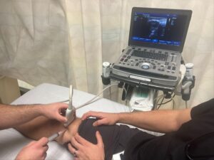
- Imaging studies: Transvaginal vs. transabdominal ultrasound (US):
- qhCG > 6000 mIU/mL => pregnancy is capable of being visualized by abdominal US
- qhCG 1500-3000 mIU/mL => pregnancy is capable of being visualized by transvaginal US
- US findings consistent with an ectopic pregnancy:
- Empty uterus (absent yolk sac and fetal pole). Note: a pseudosac may appear in the uterus similar to a gestational sac (thought to represent bleeding into the endometrial cavity by the decidual cast).2
- Adnexal mass.
- Free fluid in the cul-de-sac.
- Yolk sac and/or fetal pole in the fallopian tube.
- Fetal cardiac activity in the adnexa or adjacent peritoneum.
- Treatment:
- Unstable patient with a positive pregnancy test => Immediate OB/Gyn consult => OR
- Stable patient with ectopic pregnancy identified by US => OB/Gyn consult
- Management options vary according to the location of the ectopic pregnancy and the patient’s medical co-morbidities (e.g. methotrexate therapy is contraindicated in the setting of known blood dyscrasias, active pulmonary disease, hepatic disease, renal disease, and peptic ulcer disease, etc.).1
- Stable patient with positive pregnancy test, absent evidence of confirmed intrauterine pregnancy (IUP) (intrauterine yolk sac + fetal pole) on US => Refer for follow-up:
- Typically a repeat qhCG measurement is drawn within 48-72 hours (+/- repeat US peformed).1
- Note: Nearly 99% of normal IUPs have an increase in qhCG of > 53% within 2 days, however, this does not eliminate the possibility of an ectopic pregnancy as 13% of ectopic pregnancies will display the same appropriate rise in β-hCG.1,2
- Patients with a positive pregnancy test experiencing vaginal bleeding:
- If Rh negative and presenting within 72 hours of the onset of bleeding => administer Rhogam (standard dose: 50 μg2).
- Typically a repeat qhCG measurement is drawn within 48-72 hours (+/- repeat US peformed).1
- Pearls:
- Consider PID, ovarian torsion, threatened or incomplete abortion, appendicitis, etc. in the differential diagnosis of ectopic pregnancy.
- Maintain a high suspicion for heterotopic pregnancy in patients having undergone reproductive therapy (heterotopic pregnancy occurs in 0.3-1% of patients in this population).2
References:
- Huynh T and Patel N. Ectopic Pregnancy. In Ferri’s Clinical Advisor 2018. Philadelphia, Elsevier Saunders. 2018; 430-431.e1.
- Cline M and Young N. Pregnancy and Antepartum Care. In Conn’s Current Therapy. Philadelphia, Elsevier. 2017; 18:1097-1113.
For Additional Reading:
http://www.emdocs.net/quick-hit-ultrasound-probe-ectopic-pregnancy/