Author: Rachel Bridwell, MD (@rebridwell, EM Resident Physician, San Antonio, TX) // Reviewed by: Brit Long, MD (@long_brit, EM Attending Physician, San Antonio, TX); Alex Koyfman, MD (@EMHighAK, EM Attending Physician, UTSW / Parkland Memorial Hospital)
Welcome to EM@3AM, an emDOCs series designed to foster your working knowledge by providing an expedited review of clinical basics. We’ll keep it short, while you keep that EM brain sharp.
A 32-year-old female with a previous medical history of acne on isotretinoin presents to the ED for 2 months of headaches that awake her from sleep, more prominent in the morning. She also endorses nausea and vision changes. She notes that her vision is blurry especially in her periphery. The patient denies the possibility of being pregnant, as she is currently on OCPs. She denies fevers, neck stiffness, facial weakness, dysarthria, and personal history of cancer or venous thromboembolic events.
Vital signs (VS) include BP 188/110, HR 112, T 98.8 Oral, RR 14, SpO2 99% on room air. Neurologic exam including mental status, cranial nerves, motor, sensation, gait, and reflexes is normal.
What’s the next step in your evaluation and treatment?
Answer: Papilledema
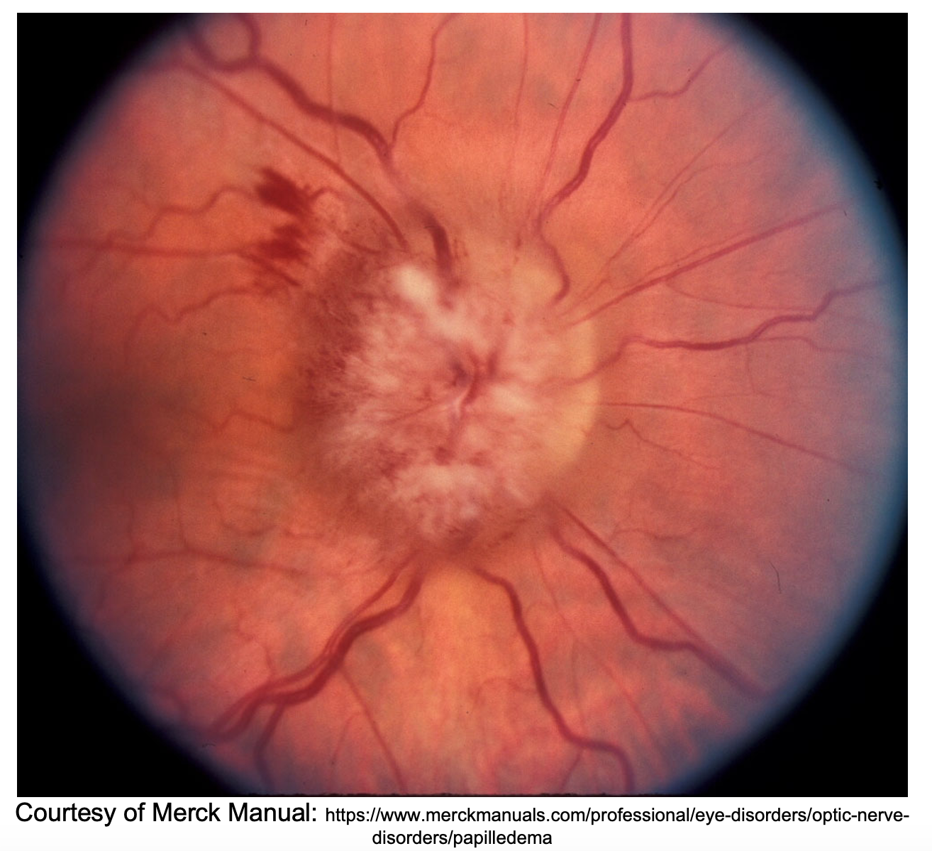
Etiology: Papilledema is the optic nerve disc swelling associated with increased intracranial pressure (ICP)1
- Results from impaired axoplasmic flow2
- Can occur secondary to many conditions
- Papilledema may lag behind intracranial hypertension
- In patients with chronic intracranial hypertension, only 6% did not demonstrate papilledema3
- Pseudopapilledema: Optic nerve edema caused by anything other than increasing ICP4
- Optic disc drusen—calcium within optic disc causes shadowing (Wall Echo Shadow [WES] sign of the eye ball)

- Inflammation of optic nerve from usually autoimmune conditions (e.g. Sarcoidosis, SLE), often unilateral.
Associated symptoms with elevated ICP:5
- Headache
- Ataxia
- Limb weakness
- Seizures
- Leptomeningeal carcinomatosis may present with numb chin syndrome6
- A sensory neuropathy primarily affecting the chin from tumor burden in proximity to branches of trigeminal nerve, especially mental and mandibular nerves7
- Fever
- Irritability or personality changes
Differential Diagnosis5:
- Idiopathic Intracranial Hypertension (IIH)
- Subdural hemorrhage/hygroma/empyema
- Subarachnoid Hemorrhage8
- Intracranial masses (primary CNS tumor and metastases)
- Meningeal disease
- Meningioma
- Leptomeningeal carcinomatosis
- ALL9
- Encephalitis
- Cavernous Venous Sinus Thrombosis (CVST)
- Progressive optic canal stenosis10
- Chiari malformations
Evaluation:
- Assess ABCs and obtain VS.
- Perform a thorough history: Question specifically timing of headaches, associated visual changes, neurological changes, current medications, prolonged prophylactic antibiotics
- Perform a physical exam:
- Fundoscopic exam is crucial, papilledema may be the only physical exam finding: can be bilateral, unilateral, asymmetric
- Can have increased OP without papilledema and normal OP with papilledema though rare11
- Fundoscopic exam is crucial, papilledema may be the only physical exam finding: can be bilateral, unilateral, asymmetric

- Pseudopapilledema: Optic nerve drusen anomalies with concomitant HA
- Assess for other CN palsies especially in CVST
- Imaging
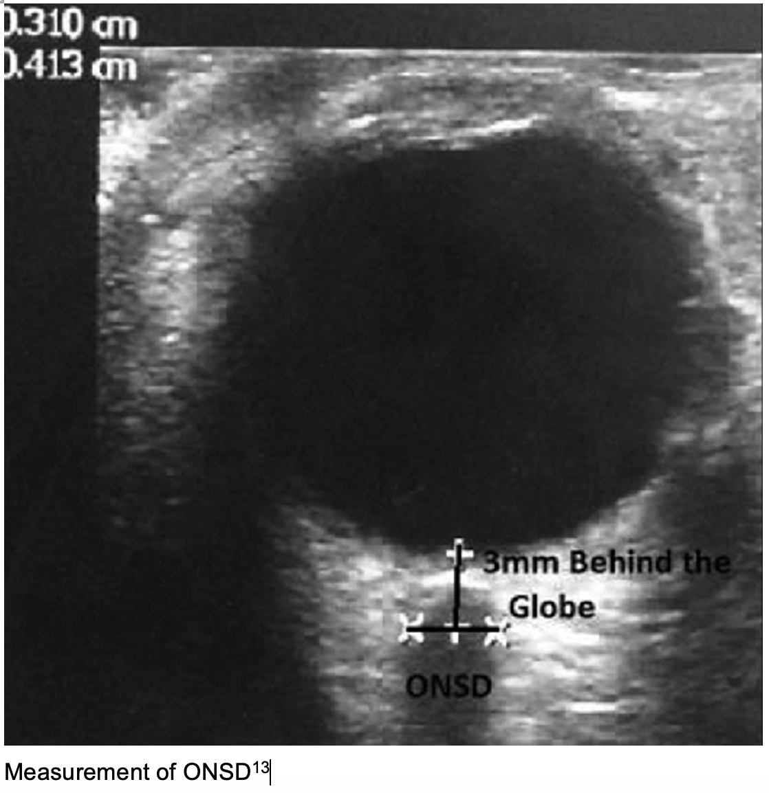
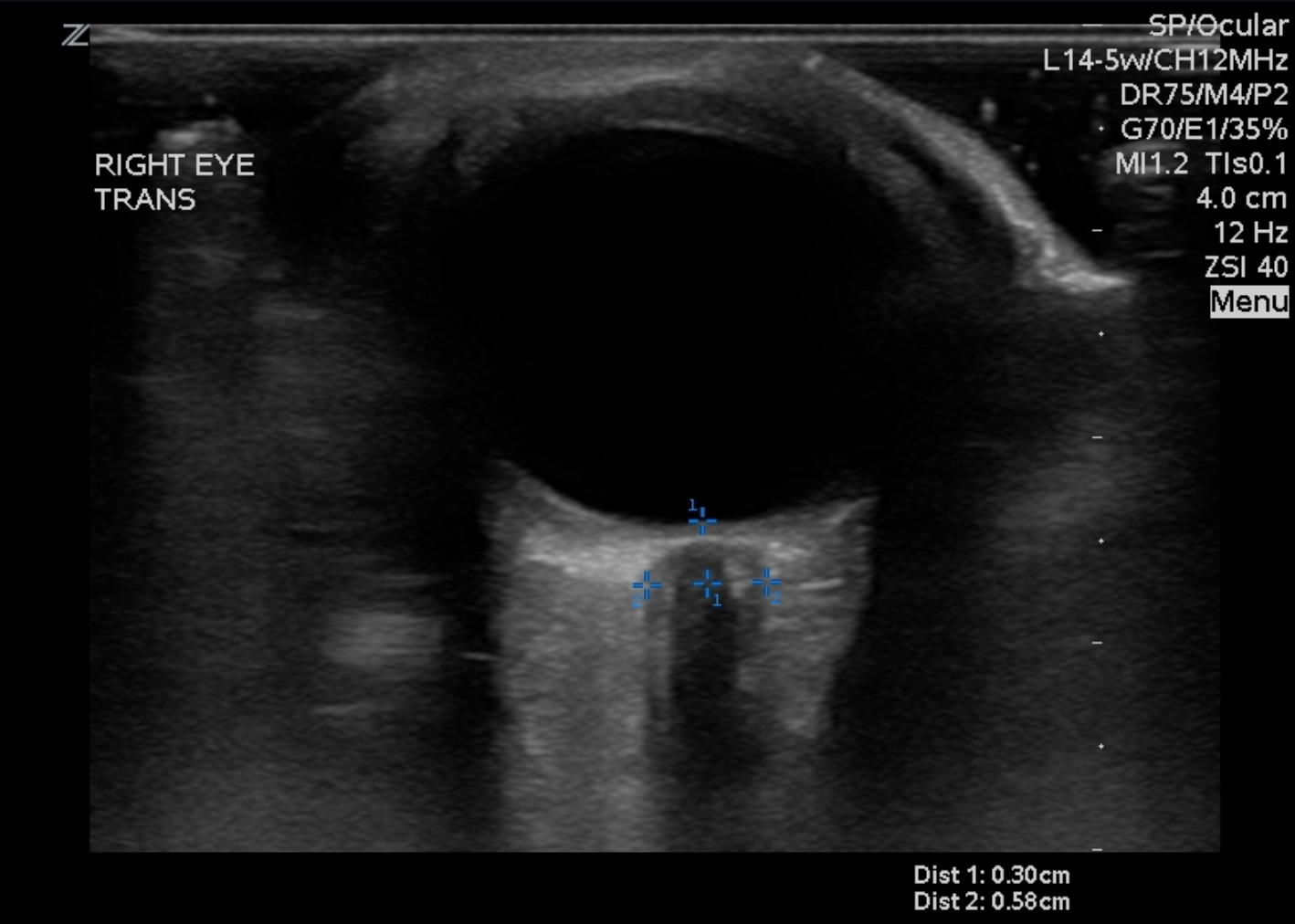
- Ocular Ultrasound to assess increased ICP
- 3mm posterior to globe, optic nerve sheath diameter>5mm increased ICP12
- Sensitivity 95.6%, Specificity 92.3%12
- 3mm posterior to globe, optic nerve sheath diameter>5mm increased ICP12


- CT can be utilized to assess for cerebral edema and mass lesion but may be normal (IIH)
- MRI5: T2 Flair can measure ONSD
- Flattening of posterior sclera, protrusion of optic papilla into globe
- MR venogram (MRV) can assess for elusive venous sinus thrombosis
- If concerned for infection or IIH, perform an LP
- LP in lateral decubitus position will show an opening pressure>25 cm H2O in IIH
- Ocular Ultrasound to assess increased ICP
Treatment:
- Stabilize ABCs
- Address any increased ICP requiring hypertonic saline or mannitol
- Treat underlying cause
- IIH:
- Acetazolamide 500 BID to treat papilledema
- Weight loss and acetazolamide together safe and effective2
- Serial LPs can be performed in acute setting but not recommended long term treatment despite chronically elevated OP14
- If fail medical management, optic nerve fenestrationà decreased HA immediately s/p surgery despite OP continually elevated6
- LP shunt less common
- Venous sinus thrombosis:
- Anticoagulation with heparin
- Admission to stroke service
- Encephalitis:
- Antibiotics
- Malignancies
- Oncology and/or hematology consult as needed
- IIH:
Disposition:
- Admit
- Consult appropriate service
Pearls:
- Keep in mind the variety of causes, and consider the differential of increased ICP. Ingestion of polar bear liver can cause IIH via Vitaminosis A, which contains 18,000 IU/g15
- Focused history and exam are essential, specifically neurologic and ophthalmologic exams.
- US is a helpful bedside evaluation and is reliable for diagnosis of papilledema.
- Management includes acute lowering of the elevated ICP and treating the underlying cause.

A 37-year-old woman presents to the emergency department for worsening chronic headache. Results of her fundoscopic exam are shown above. She has a normal noncontrast head CT. Which of the following is most likely associated with this presentation?
A) Aura preceding headache
B) Jaw claudication
C) Left ventricular hypertrophy
D) Lumbar puncture with opening pressure 31 cm H2O
Answer: D
Idiopathic intracranial hypertension, previously known as pseudotumor cerebri, typically presents as a persistent headache in an obese woman of childbearing age who is found to have papilledema on examination with an otherwise normal neurologic exam. Patients may also have visual disturbances and pulsatile tinnitus. Papilledema presents as elevation of the optic disk with blurring of the disk margins on fundoscopic exam. Retinal venous engorgement may be seen as well. All patients with papilledema should receive neuroimaging with either a head CT or brain magnetic resonance imaging to rule out mass lesions, however, patients with idiopathic intracranial hypertension will typically have normal neuroimaging. A lumbar puncture should be performed in the lateral decubitus position when the diagnosis is suspected. Opening pressures will be elevated, > 25 cm H2O. First-line treatment is with acetazolamide, to decrease cerebrospinal fluid production, and weight loss should be encouraged.

Aura preceding headache (A) is associated with a migraine. While migraines are common in young women, and patients will have normal neuroimaging if it is done, the retinal exam will also be unremarkable. A normal retinal exam will show branching dark red lines (venules) and slightly thinner bright red lines (arterioles) that converge on the optic disk. The optic disk is normally pale, with sharp, flat margins, and contains the cup, which is about half the diameter of the disk. You may be able to see the macula, which is a slightly darker area of the retina, lateral to the disk. Jaw claudication (B) is associated with temporal arteritis and can result in central retinal artery occlusion. Fundoscopic findings for central retinal artery occlusion include a pale fundus with a cherry red macular spot. Temporal arteritis is rare in patients younger than 50 years old. Left ventricular hypertrophy (C) is associated with chronic severe hypertension, which can cause hypertensive retinopathy. On fundoscopy, this is characterized by hard exudates, sausage-shaped venules, and narrowing of arterioles. Chronic hypertension can also cause papilledema and should be considered on the differential diagnosis, however, this is less likely to be present in this young of a patient.
Further Reading:
References:
- Killer HE, Jaggi GP, Miller NR. Papilledema revisited: Is its pathophysiology really understood? Clin Exp Ophthalmol. 2009;37(5):444-447. doi:10.1111/j.1442-9071.2009.02059.x
- Wall M, George D. Visual Loss in Pseudotumor Cerebri: Incidence and Defects Related to Visual Field Strategy. Arch Neurol. 1987;44(2):170-175. doi:10.1001/archneur.1987.00520140040015
- Trobe JD. Papilledema: The vexing issues. J Neuro-Ophthalmology. 2011;31(2):175-186. doi:10.1097/WNO.0b013e31821a8b0b
- Mehrpour M, Oliaee Torshizi F, Esmaeeli S, Taghipour S, Abdollahi S. Optic nerve sonography in the diagnostic evaluation of pseudopapilledema and raised intracranial pressure: A cross-sectional study. Neurol Res Int. 2015;2015. doi:10.1155/2015/146059
- Passi N, Degnan AJ, Levy LM. MR imaging of papilledema and visual pathways: Effects of increased intracranial pressure and pathophysiologic mechanisms. Am J Neuroradiol. 2013;34(5):919-924. doi:10.3174/ajnr.A3022
- Riesgo VJ, Delgado SR, Poveda J, Rammohan K. Numb chin syndrome secondary to leptomeningeal carcinomatosis from gastric adenocarcinoma. J Gastrointest Oncol. 2015;6(2):E16-E20. doi:10.3978/j.issn.2078-6891.2014.076
- Bustamante CE, Martin KD, Colyer E. Chin Numbness as a Presenting Symptom of Malignancy. R I Med J (2013). 2021;104(3):51-52. http://www.ncbi.nlm.nih.gov/pubmed/33789411. Accessed April 15, 2021.
- Perry JJ, Sivilotti MLA, Sutherland J, et al. Validation of the Ottawa Subarachnoid Hemorrhage Rule in patients with acute headache. CMAJ. 2017;189(45):E1379-E1385. doi:10.1503/cmaj.170072
- Clarke RT, Van Den Bruel A, Bankhead C, Mitchell CD, Phillips B, Thompson MJ. Clinical presentation of childhood leukaemia: A systematic review and meta-analysis. Arch Dis Child. 2016;101(10):894-901. doi:10.1136/archdischild-2016-311251
- Wright M, Miller NR, McFadzean RM, et al. Papilloedema, a complication of progressive diaphyseal dysplasia: A series of three case reports. Br J Ophthalmol. 1998;82(9):1042-1048. doi:10.1136/bjo.82.9.1042
- Spence JD, Amacher AL, Willis NR. Benign intracranial hypertension without papilledema: Role of 24-hour cerebrospinal fluid pressure monitoring in diagnosis and management. Neurosurgery. 1980;7(4):326-336. doi:10.1227/00006123-198010000-00004
- Ohle R, McIsaac SM, Woo MY, Perry JJ. Sonography of the optic nerve sheath diameter for detection of raised intracranial pressure compared to computed tomography: A systematic review and meta-analysis. J Ultrasound Med. 2015;34(7):1285-1294. doi:10.7863/ultra.34.7.1285
- Shirodkar CG, Rao SM, Mutkule DP, Harde YR, Venkategowda PM, Mahesh MU. Optic nerve sheath diameter as a marker for evaluation and prognostication of intracranial pressure in Indian patients: An observational study. Indian J Crit Care Med. 2014;18(11):728-734. doi:10.4103/0972-5229.144015
- Wall M, Kupersmith MJ, Kieburtz KD, et al. The idiopathic intracranial hypertension treatment trial clinical profile at baseline. JAMA Neurol. 2014;71(6):693-701. doi:10.1001/jamaneurol.2014.133
- Rodahl K, Moore T. The vitamin A content and toxicity of bear and seal liver. Biochem J. 1943;37(2):166-168. doi:10.1042/bj0370166


