Authors: Isabelle Piazza, MD (@isabelle_isii, EM Resident Physician, Papa Giovanni XXIII Hospital, Bergamo/University of Milan – Italy) and Roberto Cosentini, MD (@rob_cosentini, EM Attending Physician, Papa Giovanni XXIII Hospital, Bergamo- Italy) // Reviewed by: Jay Khadpe, MD; Alex Koyfman, MD (@EMHighAK)
Case
A previously healthy 77-year-old male presents with dyspnea, palpitations and fatigue and a SpO2 of 92%. The ECG shows atrial flutter with 4:1/3:1 conduction. The chest X-ray demonstrates an enlarged cardiac silhouette with retro-cardiac reduction of lung transparency and a left pleural effusion. A transthoracic echocardiogram (TTE) displays an ejection fraction (EF) of 30%.
The patient was promptly treated with IV diuretics and O2 supplementation.
Background
Heart failure (HF) is a clinical syndrome that results from any structural or functional impairment of ventricular filling or ejection of blood. The cardinal manifestations are dyspnea and fatigue, which may limit exercise tolerance, and fluid retention, which may lead to pulmonary, splanchnic congestion, and/or peripheral edema. There is not a single diagnostic test for HF because it is largely a clinical diagnosis based on a careful history and physical examination. In the emergency setting, it may be difficult to differentiate HF syndrome from other conditions that may present with a very similar clinical picture.
HF Mimics
- Pneumonia
- Chronic obstructive pulmonary disease (COPD)
- Asthma
- Pulmonary embolism
- Acute Kidney Injury (AKI)
Criteria to identify an alternative diagnosis in patients with HF symptoms
1. Consideration of conditions that mimic HF is important: The following table summarizes the most relevant demographic and epidemiological features that might orient the initial differential.
Epidemiology Key Points
- Knowing the epidemiology is pivotal to estimate the pre-test probability of a disease.
- In the emergency department, we need to know the probability of a specific diagnosis according to gender, age group, ethnicity, geographical area, social and occupational history of the patient.
- Viral and bacterial infections are a major cause of lung disease and heart failure exacerbations.
- Always bear in mind specific populations portend specific risk factors (e.g. cancer, pregnancy, immobility).
2. Risk factors: The second step is to consider different personal risk factors that may refine the pretest probability of diagnosis.
Risk factor key points
- Alcohol and smoking are important risk factors for all of the diseases included in this differential.
- Occupational history may be helpful to support specific diagnoses.
- Pay attention to the possible triggers: infections, exposure to toxins or drugs (e.g. bronchospasm after taking aspirin or NSAIDs)
3. Clinical history and presentation: The third step is recognition of typical and pathognomonic clinical presentations that can immediately rule in or rule out conditions.
Clinical history key points
- Sometimes the distinction between COPD and asthma is unclear and this may be referred to as asthma-COPD overlap syndrome.
- Unlike airflow obstruction in COPD, airflow obstruction in asthma is completely reversible in most people, either spontaneously or with treatment.
- In case of pulmonary embolism, dyspnea and/or chest pain typically have a sudden onset, which is not always the case in COPD, pneumonia, or HF
- In acute kidney injury, pulmonary congestion and/or chest pain are usually associated with severe hypertension and/or hypervolemia
4. Physical examination: The fourth step is physical exam. The table discusses the findings with the highest discriminatory power.
Physical Exam Key Points
- During physical examination, asymmetrical chest expansion, diminished breath sounds, bronchophony, and tactile fremitus can be used in combination to accurately diagnose pneumonia and pleural effusion.
- Respiratory distress correlates with severity of illness more commonly in COPD exacerbations and acute cardiogenic pulmonary edema.Distress in asthmatic patients is an ominous sign of impending respiratory arrest.
- Rhonchi are the expression of accumulation of white blood cells, fluid, and proteins in the alveolar space usually due to infection.
- Inspiratory crackles, diminished breath sounds, and cardiac dullness have high diagnostic value for advanced obstructive airway disease.
- No physical sign performs with a high degree of accuracy for diagnosing early-stage chronic obstructive pulmonary disease.
- In acute kidney disease with severe metabolic acidosis, a deep and labored breathing pattern (Kussmaul breathing) may be present.
- Congestive heart failure can be differentiated from lung disease by the presence of jugular distention (low specificity) and presence of a third heart sound (high specificity).
5. ECG findings: The fifth step is the ECG, which isnonspecific but cheap. Always worth having a look as may display typical signs that corroborate specific diagnosis.
ECG Key Points
- S1Q3T3, new onset right axis deviation, and right bundle branch block (RBBB) are highly specific for pulmonary embolism.
- Left bundle branch block (LBBB) is very often associated with left heart disease.
- New onset atrial flutter or atrial tachycardia are usually associated with pneumonia or cor pulmonale.
- Atrial fibrillation and frequent premature contractions may be present in patients chronically treated with ß2-agonists.
- Acute ECG changes may occur in electrolyte disturbances (e.g. acute kidney disease).
6. Imaging: The sixth step is imaging. Several findings on imaging can assist.
Imaging Key Points
- In the acute setting, ultrasound is the first-choice bed-side diagnostic tool.
- Echocardiography is mandatory to evaluate the ventricular function and the inferior vena cava respiratory excursion.
- Chest X-ray is a low-cost exam that allows to exclude major conditions (typical pneumonia and pulmonary edema).
- In patients with asthma, the primary role of imaging is to detect complications rather than to help make the diagnosis.
7. Laboratory testing: Last but not least is laboratory analysis. In the following table, patterns of typical lab findings are categorized according to the different alternative diagnoses.
Laboratory Test Key Points
- For the diagnosis of CHF, both BNP and the biologically inactive NT-proBNP have similar accuracy.
- One major issue is that a large number of other etiologies can elevate natriuretic peptide levels:
- coronary syndromes
- valvular heart disease
- pericardial disease
- atrial fibrillation
- cardiac surgery
- cardioversion
- older age
- anemia
- renal failure
- pulmonary hypertension
- critical illness
- sepsis
- burns
- Elevated BMI actually decreases natriuretic levels.
- Angiotensin Receptor Neprilysin Inhibitor (ARNI) increases BNP levels but not NT-proBNP levels.
- D-dimer cut-off values should be adjusted for age or clinical probability.
- COPD patients have increased risk for hepatobiliary diseases and asymptomatic elevations of hepatic transaminases, which have relatively low prevalence.
- Renal complications of COPD are common especially among patients with hypoxemia and hypercapnia.
- Nasopharygneal testing for COVID-19 and urinary antigen testing for Legionella app or S. Pneumoniae are useful to identify the underlying pathogen in cases of suspected pneumonia, COPD, and asthma exacerbations.
- In general lactate is a marker of imbalance between tissue oxygen supply and demand, and consequently may indicate overt or imminent hemodynamic compromise in cases of severe pulmonary embolism
Other rare diseases that could mimic heart failure in the emergency setting include:
-Severe anemia
-Interstitial lung disease (i.e. sarcoidosis, asbestosis, hypersensitivity pneumonitis, cryptogenic organizing pneumonia)
-Chronic infections (i.e. HIV, TBC, etc.)
-Vasculitis with pulmonary involvement
-Nephrotic syndrome
-Drug reactions
-Lung cancer
-Collagen vascular diseases
-Mechanical constrictions secondary to neoplastic infiltration
Case resolution
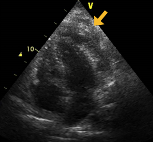
The patient didn’t improve with diuretics, and the cardiologist repeated the echo. He confirmed the reduced ejection fraction of left ventricle (30%), but with an impaired filling due to a mass adjacent to the left ventricle free wall and apex (orange arrow).
A cardiac magnetic resonance revealed a solid mass with necrotic areas sized 96 x 45 mm infiltrating the anterior-lateral and apical wall of left ventricle. A definite diagnosis of diffuse large B-cell lymphoma was made by echo-guided biopsy. After staging (Ann Arbor IV A), the patient underwent six cycles of R-COMP chemotherapy without any major side effects.
At six months follow-up, the heart failure symptoms completely resolved and the EF normalized.
In this case the HF symptoms were the first clinical presentation of a neoplastic infiltration.

Further Reading:
FOAMed
- EmDOCs “Myths in Heart Failure: Part I – ED Evaluation” http://www.emdocs.net/myths-in-heart-failure-part-i-ed-evaluation/
- EmDOCs “Myths in Heart Failure: Part II– ED Management” http://www.emdocs.net/myths-in-heart-failure-part-ii-ed-management/
- EmDOCs “All that wheezes is not asthma” – an evaluation of asthma mimics http://www.emdocs.net/wheezes-not-asthma-evaluation-asthma-mimics/
- EmDOCs “COPD Mimics: What are we missing? http://www.emdocs.net/copd-mimics-missing/
- EmDOCs “Evidence-Based Disposition of Community-Acquired Pneumonia” http://www.emdocs.net/evidence-based-disposition-of-community-acquired-pneumonia/
- EmDOCs “Acute respiratory distress syndrome: who’s at risk and ED relevant management” http://www.emdocs.net/acute-respiratory-distress-syndrome-ards-whos-risk-ed-relevant-management/
- R.E.B.E.L. EM “The Critical Pulmonary Embolism Patient” https://rebelem.com/the-critical-pulmonary-embolism-patient/
References
- Grief SN, Loza JK. Guidelines for the Evaluation and Treatment of Pneumonia. Prim Care. 2018 Sep;45(3):485-503.Musher DM, Thorner AR. Community-acquired pneumonia. N Engl J Med 2014; 371:1619.
- Ramirez JA, Wiemken TL, Peyrani P, et al. Adults Hospitalized With Pneumonia in the United States: Incidence, Epidemiology, and Mortality. Clin Infect Dis 2017; 65:1806.
- Adeloye D, Chua S, Lee C, Basquill C, Papana A, Theodoratou E, Nair H, Gasevic D, Sridhar D, Campbell H, Chan KY, Sheikh A, Rudan I; Global Health Epidemiology Reference Group (GHERG). Global and regional estimates of COPD prevalence: Systematic review and meta-analysis. J Glob Health. 2015;5:020415.
- GBD Chronic Respiratory Disease Collaborators. Prevalence and attributable health burden of chronic respiratory diseases, 1990-2017: a systematic analysis for the Global Burden of Disease Study 2017. Lancet Respir Med. 2020 Jun;8(6):585-596.
- Montagnani A, Mathieu G, Pomero F, Bertù L, Manfellotto D, Campanini M, Fontanella A, Sposato B, Dentali F; FADOI-Epidemiological Study Group. Hospitalization and mortality for acute exacerbation of chronic obstructive pulmonary disease (COPD): an Italian population-based study. Eur Rev Med Pharmacol Sci. 2020 Jun;24(12):6899-6907. doi: 10.26355/eurrev_202006_21681.
- Torres A, Peetermans WE, Viegi G, Blasi F. Risk factors for community-acquired pneumonia in adults in Europe: a literature review. Thorax 2013; 68:1057.
- Toskala E, Kennedy DW. Asthma risk factors. Int Forum Allergy Rhinol. 2015;5 Suppl 1:S11-6.
- Loftus PA, Wise SK. Epidemiology and economic burden of asthma. Int Forum Allergy Rhinol. 2015;5 Suppl 1:S7-10.
- Engelkes M, de Ridder MA, Svensson E, Berencsi K, Prieto-Alhambra D, Lapi F, Giaquinto C, Picelli G, Boudiaf N, Albers FC, Cockle SM, Bradford ES, Suruki RY, Brusselle GG, Rijnbeek PR, Sturkenboom MC, Verhamme KM. Multinational cohort study of mortality in patients with asthma and severe asthma. Respir Med. 2020;165:105919.
- Liapikou A, Ferrer M, Polverino E, et al. Severe community-acquired pneumonia: validation of the Infectious Diseases Society of America/American Thoracic Society guidelines to predict an intensive care unit admission. Clin Infect Dis 2009; 48:377.
- Konstantinides SV, Meyer G, Becattini C, Bueno H, Geersing GJ, Harjola VP, Huisman MV, Humbert M, Jennings CS, Jiménez D, Kucher N, Lang IM, Lankeit M, Lorusso R, Mazzolai L, Meneveau N, Ní Áinle F, Prandoni P, Pruszczyk P, Righini M, Torbicki A, Van Belle E, Zamorano JL; ESC Scientific Document Group. 2019 ESC Guidelines for the diagnosis and management of acute pulmonary embolism developed in collaboration with the European Respiratory Society (ERS). Eur Heart J. 2020;41:543-603.
- Ronco C, Bellomo R, Kellum JA. Acute kidney injury. 2019 Nov 23;394:1949-1964
- Aniort J, Heng AÉ, Deteix P, Souweine B, Lautrette A. Acute renal failure. 2005;365:417-30.
- Yancy CW, Jessup M, Bozkurt B, Butler J, Casey DE Jr, Drazner MH, Fonarow GC, Geraci SA, Horwich T, Januzzi JL, Johnson MR, Kasper EK, Levy WC, Masoudi FA, McBride PE, McMurray JJ, Mitchell JE, Peterson PN, Riegel B, Sam F, Stevenson LW, Tang WH, Tsai EJ, Wilkoff BL; American College of Cardiology Foundation; American Heart Association Task Force on Practice Guidelines. 2013 ACCF/AHA guideline for the management of heart failure: a report of the American College of Cardiology Foundation/American Heart Association Task Force on Practice Guidelines. J Am Coll Cardiol. 2013;62:e147-239.
- Mannino DM, Buist AS. Global burden of COPD: risk factors, prevalence, and future trends. Lancet. 2007;370:765-73.
- Cuddy R, Li G. The role of alcohol in asthma: a review of clinical and experimental studies. Am J Emerg Med. 2001;19:501-3.
- Papi A, Brightling C, Pedersen SE, Reddel HK. Asthma. 2018;391:783-800.
- Moore M, Stuart B, Little P, et al. Predictors of pneumonia in lower respiratory tract infections: 3C prospective cough complication cohort study. Eur Respir J 2017; 50.
- Shellenberger RA, Balakrishnan B, Avula S, Ebel A, Shaik S. Diagnostic value of the physical examination in patients with dyspnea. Cleve Clin J Med. 2017;84:943-950.
- Alhamed Alduihi F. ECG Abnormalities in Patients with Acute Exacerbation of Bronchiectasis and Factors Associated with High Probability of Abnormality. Pulm Med. 2021;2021:6649572.
- Glezen WP. Asthma, influenza, and vaccination. J Allergy Clin Immunol. 2006;118:1199-206
- Kameda T, Mizuma Y, Taniguchi H, Fujita M, Taniguchi N. Point-of-care lung ultrasound for the assessment of pneumonia: a narrative review in the COVID-19 era. J Med Ultrason (2001). 2021;48:31-43.
- Sheikh K, Coxson HO, Parraga G. This is what COPD looks like. Respirology. 2016;21:224-36.
- Richards JC, Lynch D, Koelsch T, Dyer D. Imaging of Asthma. Immunol Allergy Clin North Am. 2016;36:529-45
- Farrow C, King G. SPECT Ventilation Imaging in Asthma. Semin Nucl Med. 2019;49:11-15.
- Staub LJ, Mazzali Biscaro RR, Kaszubowski E, Maurici R. Lung Ultrasound for the Emergency Diagnosis of Pneumonia, Acute Heart Failure, and Exacerbations of Chronic Obstructive Pulmonary Disease/Asthma in Adults: A Systematic Review and Meta-analysis. J Emerg Med. 2019;56:53-69.
- Eibel R, Herzog P, Dietrich O, Rieger C, Ostermann H, Reiser M, Schoenberg S. Radiologe. Magnetic resonance imaging in the evaluation of pneumonia. 2006;46:267-70, 272-4.
- Fermont JM, Masconi KL, Jensen MT, Ferrari R, Di Lorenzo VAP, Marott JM, Schuetz P, Watz H, Waschki B, Müllerova H, Polkey MI, Wilkinson IB, Wood AM. Biomarkers and clinical outcomes in COPD: a systematic review and meta-analysis. 2019;74:439-446.
- Yoshizawa T, Okada K, Furuichi S, Ishiguro T, Yoshizawa A, Akahoshi T, Gon Y, Akashiba T, Hosokawa Y, Hashimoto S. Prevalence of chronic kidney diseases in patients with chronic obstructive pulmonary disease: assessment based on glomerular filtration rate estimated from creatinine and cystatin C levels. Prevalence of chronic kidney diseases in patients with chronic obstructive pulmonary disease: assessment based on glomerular filtration rate estimated from creatinine and cystatin C levels. Int J Chron Obstruct Pulmon Dis 2015;10:1283-9.
- Mapel D. Renal and hepatobiliary dysfunction in chronic obstructive pulmonary disease. Curr Opin Pulm Med. 2014;20:186-93.
- Kunc P, Fabry J, Lucanska M, Pecova R. Biomarkers of Bronchial Asthma. Physiol Res. 2020 Mar;69:S29-S34.
- Ray P, Delerme S, Jourdain P, Chenevier-Gobeaux C. Differential diagnosis of acute dyspnea: the value of B natriuretic peptides in the emergency department. QJM. 2008;101:831-43
- Galliazzo S, Nigro O, Bertù L, Guasti L, Grandi AM, Ageno W, Dentali F. Prognostic role of neutrophils to lymphocytes ratio in patients with acute pulmonary embolism: a systematic review and meta-analysis of the literature. Intern Emerg Med. 2018 Jun;13(4):603-608. doi: 10.1007/s11739-018-1805-2.
- Li J, Ye H, Zhao L. B-type natriuretic peptide in predicting the severity of community-acquired pneumonia. World J Emerg Med. 2015;6(2):131-6. doi: 10.5847wjem.j.1920-8642.2015.02.008.
- Annane D, Renault A, Brun-Buisson C, et al. Hydrocortisone plus Fludrocortisone for Adults with Septic Shock. N Engl J Med 2018; 378:809.
- Terry PD, Dhand R. Inhalation Therapy for Stable COPD: 20 Years of GOLD Reports. Adv Ther. 2020;37:1812-1828.
- Eurich DT, Marrie TJ, Minhas-Sandhu JK, Majumdar SR. Risk of heart failure after community acquired pneumonia: prospective controlled study with 10 years of follow-up. BMJ 2017; 356:j413.
- Rey JR, Caro-Codón J, Rosillo SO, Iniesta ÁM, Castrejón-Castrejón S, Marco-Clement I, Martín-Polo L, Merino-Argos C, Rodríguez-Sotelo L, García-Veas JM, Martínez-Marín LA, Martínez-Cossiani M, Buño A, Gonzalez-Valle L, Herrero A, López-Sendón JL, Merino JL; CARD-COVID Investigators. Heart failure in COVID-19 patients: prevalence, incidence and prognostic implications. Eur J Heart Fail. 2020;22:2205-2215.
- Torres A, Blasi F, Dartois N, Akova M. Which individuals are at increased risk of pneumococcal disease and why? Impact of COPD, asthma, smoking, diabetes, and/or chronic heart disease on community-acquired pneumonia and invasive pneumococcal disease. 2015;70:984-9.


