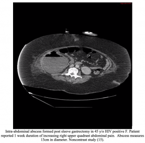
Authors: Alex Gwynne, DO (Resident Physician, San Antonio, TX) and Lloyd Tannenbaum, MD (EM Resident Physician, San Antonio, TX) // Edited by: Alex Koyfman, MD (@EMHighAK) and Brit Long, MD (@long_brit)
Case
A 44-year-old female presents to the ED with a several day history of fever, malaise, and right upper quadrant abdominal pain. She states that the pain is a 5/10 and radiates to her right shoulder and scapula. The pain is non-positional and not worse with deep inspiration. She underwent a cholecystectomy for symptomatic cholelithiasis 2 weeks ago. After surgery, she had an uncomplicated course, but 4 days ago she started to develop a low-grade fever, chills, RUQ pain, and generalized malaise. The patient is ill appearing and diaphoretic.
Abdominal exam reveals laparoscopic dry and well-healing surgical incisions with no signs of infection. Abdominal auscultation reveals regular bowel sounds. The abdomen is diffusely tender throughout, but worse in the RUQ. Vital signs include a blood pressure of 102/74, heart rate of 94, respiratory rate of 20, and an O2 sat of 96% on room air. Her oral temperature is 101.4 degrees Fahrenheit.
During the patients stay in the ED, her blood pressure begins to fall, and she become progressively more tachycardic and tachypneic. As you work on resuscitating her, you wonder what could be the cause of these symptoms and how you should direct your interventions.
Introduction/Etiology
Intra-abdominal abscesses are a rare surgical complication and may not be at the forefront of the emergency physician’s mind when evaluating a patient with abdominal pain. With the myriad of causes of abdominal pain, a focused history, physical, and strong clinical suspicion are key to making this unusual diagnosis. Vague and varied patient presentations make prompt diagnosis difficult (1,2). These patients can quickly develop bacteremia and subsequently progress to sepsis with shock requiring intubation and vasopressors (3). Progression of this disease process leads to increased morbidity and mortality, as well as significant costs with many patients requiring lengthy intensive care unit stays (2).
Definition
An intra-abdominal abscess is a collection of pus or infected material and is usually due to a localized infection inside the peritoneal cavity. It can involve any intra-abdominal organ or can be located freely within the abdominal or pelvic cavities, including in between loops of bowel (2).
Intra-abdominal abscesses occur almost exclusively secondary to preexisting disease processes and often involve multiple bacterial, fungal, or parasitic infectious agents (2). They usually are a secondary complication of intra-abdominal pathology, with several more common causes including perforation of a viscus, appendicitis, or diverticulitis; gangrenous cholecystitis; mesenteric ischemia with bowel infection; and necrotic pancreatitis (3,9). Less common causes included penetrating abdominal trauma, inflammatory conditions such as Crohn’s disease, malignancy, or postoperative causes (2,3,9). Post-operative abscesses generally present 4-21 days after surgery and are caused most frequently by preoperative contamination, spillage of bowel contents during surgery, or postoperative anastomotic leaks (5).
Differential
Patients will present with a range of many different symptoms, primarily based on the size and location of the abscess. Some patients will present with only vague abdominal pain while others will come in with fulminant septic shock. Due to this large spectrum of presentation, it is important to keep a broad differential:
Appendicitis
Cholecystitis
Pancreatitis
Diverticulitis
Bowel Obstruction
Bowel Perforation
Mesenteric Ischemia
Kidney stone
Gastritis
Gastroenteritis
AAA
Ruptured gastric ulcer
Malignancy
Cystitis
Pyelonephritis
Presentation/Evaluation
Physical exam findings can vary greatly. Presentation can range from mild abdominal pain and fever to full on septic shock. Abscesses can develop anywhere in the abdomen and retroperitoneum depending on what the causative factor is, thus tenderness throughout any abdominal quadrant can be seen as well. A palpable mass is an uncommon finding that depends mostly on the size and location of the abscess, as well as the body habitus of the patient (2,3).
Laboratory testing is not specific. A CBC showing a leukocytosis >20,000 or a left shift can point towards abscess formation; however, a normal WBC count or lack of fever does not exclude the diagnosis. The elderly or immunosuppressed may not be able to mount a reactive leukocytosis or fever (2,4,11). An ESR or CRP may also be elevated in cases of intra-abdominal abscess, but again are not specific (2). Blood cultures have limited value, as bacteremia related to the organisms present in an infected abdominal site range from 0% to 5% in an appendicitis study and percutaneous drainage study, respectively (12,13). As most abscesses are caused by many different types of bacteria, blood cultures often reveal polymicrobial bacteremia. Interestingly, more than 90% of intra-abdominal abscesses contain anaerobic organisms, particularly Bacteroides fragilis. Thus, postoperative patients presenting with Bacteroides bacteremia suggests an intra-abdominal source of their sepsis (2).
Imaging is the most definitive tool for diagnosing an intra-abdominal abscess. Ultrasound, in the hands of a skilled practitioner, has a sensitivity ranging 71% to 97% (2,14). Ultrasound is convenient, widely available, quick, cheap, and does not dose the patient with any radiation. Its drawbacks are that it may be obstructed by surgical dressings, and images may be limited due to body habitus. CT scan is the definitive option for the evaluation of intra-abdominal abscess (7). It has a greater than 95% accuracy in diagnosing intra-abdominal abscesses and is not hindered by post-surgical dressings or body habitus (3). IV contrast can be used to help differentiate abscess from surrounding structures and to help determine the ability for percutaneous drainage. Oral contrast can assist in the differentiation of a fluid-filled extraluminal structure from normal intestine, while oral contrast extravasation indicates a fistula or anastomotic leak. To our knowledge, there have not been any trials directly comparing the diagnostic sensitivity of a CT scan with IV and PO contrast vs IV only in patients with intra-abdominal abscess. Based on the American College of Radiology Appropriateness Criteria for post-operative patients with acute non-localizing abdominal pain, we recommend obtaining a CT scan with IV contrast (16). In post-surgical patients, discussion with surgery is recommended to determine the best imaging modality. Since there are no trials comparing these two studies head to head, and given how sick these patients can present, it is unlikely that the patient would be able to tolerate PO contrast.


Treatment
Once identified, treatment of intra-abdominal abscesses follows three main tenants: resuscitation (mainly for patients presenting in sepsis/septic shock), source control, and antimicrobial therapy (5).
ED physicians should discuss drainage options with the surgical service. If signs of hemodynamic instability are present, patients should be volume resuscitated with IV fluids and, if necessary, pressor support (1). Blood cultures should be obtained, and broad-spectrum IV antibiotics should be promptly administered. In patients with sepsis or septic shock, IV antibiotics with broad-spectrum coverage should be initiated immediately as outcome worsens with each hour delay of antimicrobial therapy (8). The patient should be admitted for intravenous antibiotics and further workup.
Antibiotic therapy should promptly be initiated in the ED as soon as a diagnosis of intra-abdominal abscess is made or considered likely, including patients who are not currently showing signs of septic shock (1). Empiric antibiotic therapy should be initiated before abscess drainage and should be concluded once all signs of sepsis have resolved (3). Infections are broken down into community acquired, community acquired high risk, and hospital acquired. This differentiation is often difficult in the ED. Discussion with the surgical consult for antimicrobials is recommended, with broad-spectrum coverage including piperacillin/tazobactam (4.5 g intravenously every 6 hours) and vancomycin (15-20 mg/kg intravenously every 8-12 hours) to cover pseudomonal strains, MRSA, and enterococcus (2). Cefepime (2 g IV every 8 hours) and metronidazole (500 mg intravenously every 8-12 hours) can be used in place of piperacillin/tazobactam. In patients who are immunosuppressed, candida species may play an important pathogenic role, and treatment with fluconazole (400-800 mg/day intravenously) may be indicated (2,3).
Once the source has been identified via imaging, source control is achieved through abscess drainage. Nearly all intra-abdominal abscesses require drainage, either by laparoscopic/open surgical drainage or percutaneous methods (7). Abscesses that do not require drainage include small (< 2 cm) pericolic or periappendiceal abscesses, or those that are draining spontaneously to the skin or into the bowel (7). Percutaneous CT guided drainage has become the standard treatment of most intra-abdominal abscesses and is preferred to open drainage when feasible (3). Features of abscesses likely requiring drainage include: multiple abscesses, loculations, enteric anastomosis, infected hematoma, or those that the trajectory to reach the abscess would seed additional cavities (1,2). Cultures should be taken from the drainage. Improvement in clinical status within 3 days indicates successful drainage, whereas failure to improve could indicate additional sources of sepsis or inadequate drainage (3).
Case Conclusion
For our patient, you quickly notice the signs of septic shock, initiate fluid resuscitation, and place her on a non-rebreather at 15L. With the fluids and oxygen, her O2 sat comes up to 95%, and her blood pressure rises to 110/60. You start broad spectrum antibiotics to include Vanc and Zosyn and order a CT scan with IV contrast. The scan shows an intrabdominal abscess in her right upper quadrant. You call your surgical consultants, who come see her in the ER agree to admit her directly to the OR for a wash out. The patient then made an uneventful recovery and was discharged 3 days later.
Key points
-History is paramount in making this diagnosis quickly. Look out for red flags for abdominal abscess such as patients presenting with fever, abdominal pain, or sepsis with a history of abdominal surgery, abdominal trauma, or conditions such as pancreatitis, Crohn’s disease, diverticulitis, appendicitis, cholecystitis, or other inflammatory conditions.
-Labs are of limited value. Blood cultures may show polymicrobial bacteremia.
-Abdominal CT w/ contrast is needed for diagnosis.
-Treatment is source control via percutaneous or surgical drainage and broad-spectrum antibiotics. Surgery should be consulted early.
-Antibiotics should be given as soon as the diagnosis is made or suspected in critically ill patients. Broad-spectrum coverage is recommended.
References/Further Reading
- Joseph S. Solomkin, John E. Mazuski, John S. Bradley, et al; Diagnosis and Management of Complicated Intra-abdominal Infection in Adults and Children: Guidelines by the Surgical Infection Society and the Infectious Diseases Society of America, Clinical Infectious Diseases, Volume 50, Issue 2, 15 January 2010, Pages 133–164
- Kreiner, L., MD, FACS. (2018, January). Intra-abdominal abscess. Retrieved September 22, 2018, from https://bestpractice.bmj.com/topics/en-us/996
- Saber, A. A., MD, MS, FACS, FASMBS. (2018, August 15). Abdominal Abscess: Background, Anatomy, Pathophysiology. Retrieved September 23, 2018, from https://emedicine.medscape.com/article/1979032-overview
- Stapczynski, J. S., & Tintinalli, J. E. (2016). Tintinalli’s emergency medicine: A comprehensive study guide, 8th Edition. New York: McGraw-Hill Education.Chapter 83: Bowel obstruction.
- Stapczynski, J. S., & Tintinalli, J. E. (2016). Tintinalli’s emergency medicine: A comprehensive study guide, 8th Edition. New York: McGraw-Hill Education.Chapter 87: Complications of General Surgery Procedures.
- Walls, R. M., Hockberger, R. S., Gausche-Hill, M., & Bakes, K. M. (2018). Rosen’s emergency medicine: Concepts and clinical practice. Philadelphia, PA: Elsevier.Chapter 9: Fever in the Adult Patient.
- Ansari, P., MD. (2017, January). Intra-Abdominal Abscesses – Gastrointestinal Disorders. Retrieved September 21, 2018, from https://www.merckmanuals.com/professional/gastrointestinal-disorders/acute-abdomen-and-surgical-gastroenterology/intra-abdominal-abscesses
- Kumar A, Roberts D, Wood KE, et al. Duration of hypotension before initiation of effective antimicrobial therapy is the critical determinant of survival in human septic shock. Crit Care Med. 2006;34:1589-1596.
- Lopez N, Kobayashi L, Coimbra R. A Comprehensive review of abdominal infections. World Journal of Emergency Surgery : WJES. 2011;6:7. doi:10.1186/1749-7922-6-7.
- Muzio, B. D. (n.d.). Intra-abdominal abscess after appendectomy | Radiology Case. Retrieved September 24, 2018, from https://radiopaedia.org/cases/intra-abdominal-abscess-after-appendectomy.
- Cosse C, Regimbeau JM, Fuks D, et al: Serum procalcitonin for predicting the failure of conservative management and the need for bowel resection in patients with small bowel obstruction. J Am Coll Surg 216: 997, 2013. [PMID: 23522439]
- Cueto J, D’Allemagne B, Vazquez-Frias JA, et al. Morbidity of laparoscopic surgery for complicated appendicitis: an international study, Surg Endosc , 2006, vol. 20 (pg. 717-20).
- Akinci D, Akhan O, Ozmen MN, et al. Percutaneous drainage of 300 intraperitoneal abscesses with long-term follow-up, Cardiovasc Intervent Radiol , 2005, vol. 28 (pg. 744-50).
- Panés J, Bouzas R, Chaparro M, et al. Systematic review: the use of ultrasonography, computed tomography and magnetic resonance imaging for the diagnosis, assessment of activity and abdominal complications of Crohn’s disease. Aliment Pharmacol Ther. 2011;34:125-145.
- Kaluza, M. (n.d.). Abdominal abscess | Radiology Case. Retrieved October 1, 2018, from https://radiopaedia.org/cases/abdominal-abscess-1
- ACR Appropriateness Criteria® Acute nonlocalized abdominal pain. (n.d.). Retrieved from https://acsearch.acr.org/docs/69467/Narrative/-American College of Radiology.


2 thoughts on “Intra-abdominal Abscess – Pearls and Pitfalls”
Fantastic post! I wonder if the metronidazole would be better placed with the cefepime regimen, as this cephalosporin lacks anaerobic coverage. Pip/tazo’s spectrum of activity has historically been sufficient to cover those anaerobes as monotherapy.
Hi Marc, thanks for reading, and you are correct! This has been updated in the post to reflect this.