Author: Sarah Brubaker, MD (EM Physician, US Army) // Reviewed by: Alex Koyfman, MD (@EMHighAK) and Brit Long, MD (@long_brit)
Case 1:
A 23-year-old female with type-2 diabetes presents with a “swollen index finger.” Her cat bit her finger 4 days ago; since then, her finger has become progressively more swollen, red, and painful. The patient developed a subjective fever this morning, and her pain has become excruciating, which is why she came to the Emergency Department (ED).
The patient’s heart rate is 110, her temperature is 101.1F, and the remainder of her vital signs are normal. The patient is diaphoretic and in moderate distress due to pain. Her right index finger is diffusely swollen, with a small puncture wound on the fingertip. The patient is holding the finger in a slightly flexed position, because even minimal extension causes excruciating pain. The entire finger is tender, most prominently on the ventral aspect. The remainder of her exam is normal.
Background
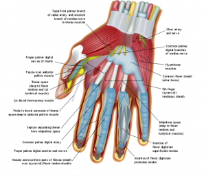
Hand infections can be intimidating for many reasons. First, the hands are vital contributors to daily life, and most people’s wellbeing and career depend on their hands. Medically, the anatomy of hands is complex, with multiple important structures in very close proximity to each other.1 In addition, these connective tissues possess relatively low vascularity, which makes them more prone to infection.2 Therefore, even seemingly benign infections can spread rapidly and lead to significant disability if not treated expeditiously. For these reasons, it is especially important to consider hand-threatening disease for every patient with a potential hand-infection who presents to the ED–even though the presentation can range from anything as simple as superficial cellulitis to necrotizing deep tissue infection.
Although the general principles of treating skin infections apply to hand infections as well, there are a few principles that are unique, and must be considered by every emergency provider (EP). The approach to hand infections can be guided by answering several questions: Is the infection superficial, or does it extend to deeper structures? Is imaging necessary? Which antibiotics are appropriate (and are there any special considerations for antibiotic coverage)? If there is an associated break in the skin (abrasion, laceration, etc.), are there any specific wound care considerations?
Question 1: Is this a superficial or deep-space infection?
Because the hand is divided into multiple compartments, and each compartment is in very close proximity to the others, superficial hand infections can rapidly spread to deeper structures. Although the most common deep-space infection is flexor tenosynovitis,1 any deep structure has the potential to become infected, including the bursae, fascial planes, potential spaces (most commonly the thenar space),3 webspaces (“collar button abscess”), joints (septic arthritis), and bones (osteomyelitis).4 All of these infections require surgical consultation, as many require intravenous antibiotics and operative incision + drainage.
Fortunately, deep-space infections in the hand are relatively rare.5 Although osteomyelitis of the hand accounts for 1 to 6% of all hand infections,6 it is almost always secondary to open fractures or surgical manipulation (e.g. open fixation). Septic arthritis is also very rare, and the cases are usually traumatic or iatrogenic.6,7 The most common cause of septic arthritis in the hand is human bite.6
It is important to consider septic arthritis in the warm, swollen, painful wrist or finger; however, arthritis, gout, and cellulitis are much more common causes of these symptoms.
The most common deep-space infection in the hand is flexor tenosynovitis (FTS),8 which is a purulent infection in the flexor tendon sheath. Although FTS can develop via hematogenous spread, it  most commonly develops as a result of a puncture wound or another injury.1 Because the flexor tendon sheaths connect in the palm, FTS can rapidly spread proximally. The classic “Kanavel signs” that indicate FTS include fusiform swelling, pain with passive extension, a finger that is passively held in flexion, and tenderness along the flexor tendon sheath. On ultrasound, fluid may be visualized within the tendon sheath.9 This ultrasound finding is up to 95% sensitive and 74.4% specific for FTS. Inflammatory markers, such as ESR and CRP, may be elevated, but are not sufficiently sensitive or specific to be relied upon.9
If labs are not useful to diagnose FTS, what can help you clench the diagnosis? Unfortunately, diagnosing deep-space infections of the hand can sometimes be tricky. Because hand infections are often associated with low bacterial load, systemic symptoms, lymphadenopathy, and other overt evidence of deep-space infection do not reliably occur.4,7 WBC and CRP are helpful in supporting the diagnosis, but they are not sensitive nor specific.10 In a study of 82 patients with presumed or surgical diagnosis of pyogenic flexor tenosynovitis, the sensitivity of elevated WBC (>11 cells/L), ESR (>15 mm/h), and CRP (>1.0 mg/dL) was found to be 39%, 41%, and 76%, respectively.11 The negative predictive value was 4%, 3%, and 13%.11 Therefore, although it is common to obtain a CBC, ESR, and CRP, use these results with caution. Imaging is also not always helpful, as CT and XR images are not reliably sensitive in detecting deep-space infections. Ultrasound is perhaps the most useful imaging modality; for FTS, look for fluid around the affected flexor tendon. In a prospective observational trial of 57 patients, this technique was 94% sensitive, 65% specific, 63% PPV, 95% NPV. 9
Ultimately, use your clinical gestalt and the overall clinical picture. If you have high clinical suspicion for deep-space infection, it is important to involve your orthopedic surgeon early. If you are unsure, it is better to have your orthopedist evaluate the patient, because even superficial infections can worsen rapidly.
Case Resolution: You perform a bedside ultrasound, which reveals fluid within the flexor tendon sheath. The WBC and the other inflammatory markers are surprisingly normal. You suspect flexor tenosynovitis. You call the orthopedic surgeon, who recommends intravenous antibiotics and will admit the patient for operative drainage.
Once you have sufficiently lowered your suspicion for deep-space infection, the principles are the same as for other soft-tissue infections, including treating cellulitis with antibiotics and performing incision + drainage on abscesses. However, there are several special considerations. The remainder of this article will discuss the management of superficial infections that can be treated and discharged from the ED.
Question 2: When is imaging appropriate?
Imaging may be helpful when attempting to distinguish between superficial and deep-space infections, as discussed above. However, beyond this context, imaging is of limited utility. Nonetheless, it is important to obtain imaging to evaluate for fractures (if the infection is related to a crush injury or other significant trauma) and retained foreign bodies (especially in animal bites). X-ray is highly sensitive for radio-opaque objects (like glass, gravel, and metal)12, but radiolucent objects (like wood and plastic) may be missed on CT and X-ray. Some studies cite the sensitivity of X-ray for detection of radiolucent objects as low as nearly 0%.13,14 Ultrasound is much more sensitive than X-ray and CT. Depending on the composition of the foreign body, ultrasound has up to 95% sensitivity in detecting soft tissue foreign bodies.15,16 An emDocs article from 2016 gives a great overview of this topic, in addition to tips for how to perform the ultrasound (http://www.emdocs.net/ultrasound-for-retained-radiolucent-foreign-body-in-soft-tissue/). Ultrasound is particularly helpful because in addition to helping evaluate for the presence of foreign bodies, it can also confirm the diagnosis of cellulitis and evaluate for potential abscess.
Question 3: What antibiotics should I use?
There are several unique considerations dictating antibiotic selection for hand infections. First, MRSA is more common in hand infections compared to other wounds.17,18 Staphylococcus is responsible for up to 84% of all hand infections;18–20 MRSA accounts for 42-80% of hand infections.17,19 The other most common organism is beta-hemolytic streptococcus, which is common skin flora. A meta-analysis in 2014 found the most common organisms in hand infections to be MRSA (49%), MSSA (21%), polymicrobial (20%), and Streptococcus pyogenes (8%).18 Based on these data, most sources recommend covering for both streptococcus and MRSA.
When an infection is caused by an animal exposure or bite, the antibiotic regimen may be greatly influenced by the mode of inoculation.7 For example, although dog bites account for up to 90% of all animal bite injuries, they are less likely to become infected than cat bites. This is probably because dogs have broader teeth and are likely to cause larger lacerations and crush injuries;21 while cats have smaller, sharper teeth that may puncture deeper than initially apparent.22 Anywhere from 30 to 76% of all cat bites become infected.2,7,23 In general, Amoxicillin-Clavulanic Acid is the antibiotic-of-choice for animal bites (including human bites). Clindamycin plus ciprofloxacin is an acceptable regimen for patients with penicillin allergies. Doxycycline is the antibiotic-of-choice for infections related to injuries from water creatures.

Question 4: Anything special about wound care?
As a whole, wound care is the same in the hand as in any other part of the body. However, if the wound crosses a joint or involves the finger, it is important to immobilize the affected area.17 In addition, warm soaks are easy to perform and may be helpful to reduce inflammation and promote proper drainage of any purulence. Lastly, because there are so many compartments and deep spaces in the hand, infections in the hand may require surgical intervention despite appropriate medical management.7 Therefore, it is generally recommended to have the patient return to the ED or see another provider in 48 hours.
Case 2:
A 22-year-old male with no medical conditions presents with the chief complaint of “swollen fingertip.” The patient says he noticed the finger was slightly painful and red 2 days ago, but since then, it has become increasingly more swollen, red, and painful. He denies fevers, chills, or any other symptoms. He does not recall any recent trauma.
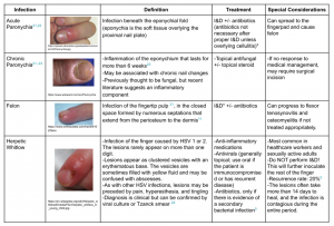
Because the fingertip is the most common site of hand infections,28 and because fingertip infections have their own unique considerations, they warrant their own discussion. The nuances to the diagnosis and treatment of these infections are beyond the scope of this paper, but the table below reviews the basics:
 *Although paronychias can be drained by simply elevating the eponychia with an 18-gauge needle or #11-blade scalpel, felons require true I&D (generally longitudinally along the lateral aspects of the finger, if possible, to avoid incising the neurovascular bundles in the finger pad) and potentially evaluation by an orthopedic surgeon.29
Conclusion: Although hand infections can be daunting, the Emergency Provider can follow the same tenets for hand infections as for any other infections. However, it is important to keep these clinical pearls in mind.
Clinical Pearls
- Use ultrasound and your physical exam to distinguish between superficial and deep-space infection. If you have high concern for deep-space infection, consult your orthopedist, regardless of lab values.
- If you discharge the patient, remember to consider MRSA coverage, soft-tissue immobilization, and 48-hour re-check. If you performed an I&D, regular warm water soaks will probably be useful to ensure proper continued drainage.
- Don’t forget to ask about recent exposures and injuries; human bites, animal bites, and other zoonotic exposures require specific antibiotic coverage.
- If there is an associated wound, check the patient’s tetanus immunization status, and give the patient a tetanus booster if it was not updated in the last 5 years.
- Make sure a paronychia is NOT herpetic whitlow, because attempting to I&D herpetic whitlow will only make matters worse.
References
- Tintinalli JE, Stephan Stapczynski J, John Ma O, Cline DM, Meckler GD. Tintinalli’s Emergency Medicine: A Comprehensive Study Guide, 8th Edition. McGraw Hill Professional; 2016.
- Kennedy SA, Stoll LE, Lauder AS. Human and Other Mammalian Bite Injuries of the Hand. Journal of the American Academy of Orthopaedic Surgeons. 2015;23(1):47-57. doi:10.5435/jaaos-23-01-47
- Osterman M, Draeger R, Stern P. Acute hand infections. J Hand Surg Am. 2014;39(8):1628-1635; quiz 1635.
- Malizos KN, Papadopoulou ZK, Ziogkou AN, et al. Infections of Deep Hand and Wrist Compartments. Microorganisms. 2020;8(6). doi:10.3390/microorganisms8060838
- Pinder R, Barlow G. Osteomyelitis of the hand. Journal of Hand Surgery (European Volume). 2016;41(4):431-440. doi:10.1177/1753193415612373
- Flevas DA, Syngouna S, Fandridis E, Tsiodras S, Mavrogenis AF. Infections of the hand: an overview. EFORT Open Rev. 2019;4(5):183-193.
- Teo WZW, Chung KC. Hand Infections. Clinics in Plastic Surgery. 2019;46(3):371-381. doi:10.1016/j.cps.2019.03.003
- Chapman T, Ilyas AM. Pyogenic Flexor Tenosynovitis: Evaluation and Treatment Strategies. J Hand Microsurg. 2019;11(3):121-126.
- Jardin E, Delord M, Aubry S, Loisel F, Obert L. Usefulness of ultrasound for the diagnosis of pyogenic flexor tenosynovitis: A prospective single-center study of 57 cases. Hand Surg Rehabil. 2018;37(2):95-98.
- Whitaker CM, Low S, Gorbachova T, Raphael JS, Williamson C. Imaging and Laboratory Workup for Hand Infections. Hand Clin. 2020;36(3):285-299.
- Bishop GB, Born T, Kakar S, Jawa A. The diagnostic accuracy of inflammatory blood markers for purulent flexor tenosynovitis. J Hand Surg Am. 2013;38(11):2208-2211.
- Tseng H-J, Hanna TN, Shuaib W, Aized M, Khosa F, Linnau KF. Imaging Foreign Bodies: Ingested, Aspirated, and Inserted. Ann Emerg Med. 2015;66(6):570-582.e5.
- Friedman DI, Forti RJ, Wall SP, Crain EF. The utility of bedside ultrasound and patient perception in detecting soft tissue foreign bodies in children. Pediatr Emerg Care. 2005;21(8):487-492.
- Manthey DE, Storrow AB, Milbourn JM, Wagner BJ. Ultrasound versus radiography in the detection of soft-tissue foreign bodies. Ann Emerg Med. 1996;28(1):7-9.
- Aras MH, Miloglu O, Barutcugil C, Kantarci M, Ozcan E, Harorli A. Comparison of the sensitivity for detecting foreign bodies among conventional plain radiography, computed tomography and ultrasonography. Dentomaxillofac Radiol. 2010;39(2):72-78.
- Tantray MD, Rather A, Manaan Q, Andleeb I, Mohammad M, Gull Y. Role of ultrasound in detection of radiolucent foreign bodies in extremities. Strategies Trauma Limb Reconstr. 2018;13(2):81-85.
- McDonald LS, Bavaro MF, Hofmeister EP, Kroonen LT. Hand Infections. The Journal of Hand Surgery. 2011;36(8):1403-1412. doi:10.1016/j.jhsa.2011.05.035
- Tosti R, Samuelsen BT, Bender S, et al. Emerging multidrug resistance of methicillin-resistant Staphylococcus aureus in hand infections. J Bone Joint Surg Am. 2014;96(18):1535-1540.
- Houshian S, Seyedipour S, Wedderkopp N. Epidemiology of bacterial hand infections. Int J Infect Dis. 2006;10(4):315-319.
- Morgan M. Hospital management of animal and human bites. J Hosp Infect. 2005;61(1):1-10.
- Benson LS, Edwards SL, Schiff AP, Williams CS, Visotsky JL. Dog and cat bites to the hand: treatment and cost assessment. J Hand Surg Am. 2006;31(3):468-473.
- Medeiros I, Saconato H. Antibiotic prophylaxis for mammalian bites. Cochrane Database Syst Rev. 2001;(2):CD001738.
- Kwo S, Agarwal JP, Meletiou S. Current treatment of cat bites to the hand and wrist. J Hand Surg Am. 2011;36(1):152-153.
- for Disease Control C, (cdc P, Others. Updated recommendations for use of tetanus toxoid, reduced diphtheria toxoid and acellular pertussis (Tdap) vaccine from the Advisory Committee on Immunization Practices, 2010. MMWR Morb Mortal Wkly Rep. 2011;60(1):13-15.
- Shoji K, Cavanaugh Z, Rodner CM. Acute fight bite. J Hand Surg Am. 2013;38(8):1612-1614.
- Akram SM, Aboobacker S. Mycobacterium Marinum. In: StatPearls. StatPearls Publishing; 2020.
- Bross MH, Soch K, Morales R, Mitchell RB. Vibrio vulnificus infection: diagnosis and treatment. Am Fam Physician. 2007;76(4):539-544.
- Barger J, Garg R, Wang F, Chen N. Fingertip Infections. Hand Clin. 2020;36(3):313-321.
- Bowen WT, Slaven EM. Evidence-Based Management Of Acute Hand Injuries In The Emergency Department.Emerg Med Pract. 2014;16(12):1-25; quiz 26-27.


