Originally published on Ultrasound G.E.L. on 4/15/19 – Visit HERE to listen to accompanying PODCAST! Reposted with permission.
Follow Dr. Michael Prats, MD (@PratsEM), Dr. Creagh Bougler, MD (@CreaghB), and Dr. Jacob Avila, MD (@UltrasoundMD) from Ultrasound G.E.L. team!
The diagnostic accuracy of ultrasonography in determining the reduction success of distal radius fractures
Turk J Emerg Med August 2018 – Pubmed Link
Take Home Points
1. Point of care ultrasound appears to accurately determine success of reduction of distal radius fractures in adults.
2. Xray offers additional valuable information and therefore will likely still be necessary in most situations.
Background
We know that ultrasound can be used for a lot of musculoskeletal complaints. In fact, many studies have shown that is is particularly accurate for fractures of the extremities. The next question is if point-of-care ultrasound can entirely replace xray for these situations. However, xrays tell us more information beyond is it broken or not. We can look for displacement, angulation, and then also guide reduction of the fracture. On the other hand, using ultrasound would be advantageous in that it can be faster and does not require radiation (pediatric doctors love that). More importantly, you can check the reduction easily before splinting the patient. This could potentially prevent the frustrating scenario of having to take down a splint, re-sedate, and re-reduce. There have been a few studies that show this can work in pediatric patients (Chinnock 2011, Ang 2010, Patel 2009, Chen 2007). This article takes a look at using ultrasound to assess for successful reduction of adult patients with distal radius fractures.
Questions
What is the accuracy of bedside US in the detection of success in reduction of distal radius fractures?
Can ultrasound detect the cause of an unsuccessful reduction?
Population
Single center in Turkey
Inclusion:
- adults > 18 years old
- Linear distal radius fracture requiring closed reduction (defined based on 2 view xray)
Exclusion:
- Did not need a reduction
- Fracture was not linear
- Did not consent
- Required operative reduction
Design
Prospective cross sectional convenience sample
Patient enrolled. Prereduction ultrasound performed (a single EM resident did all the scans).
Fracture reduced (by orthopedic resident). Post-reduction ultrasound performed (but not disclosed to orthopedic resident). This ultrasound later interpreted by a single attending EM physician as successful or not. Cast placed.
Post reduction xray performed. Orthopedic resident could choose to perform further reduction but it appears as though these patients would not receive a second post-reduction ultrasound in those cases. Accuracy of post-reduction ultrasound compared to “gold standard” of orthopedic hand surgeon interpretation (who was not involved in patient care) of the post-reduction xray. Note that this occurred at a later time.
Considered double-blinded because the EM attending who determined the ultrasound results did not see the patient or the xray images and the Orthopedic attending who determined the xray results did not see the patient or ultrasound images.
Who did the ultrasounds?
EM resident trained in extremity ultrasound, 2 years of experience. Had scanned at least 25 patients under supervision.
The Scan
Linear Probe

Two view
- Dorsal side of radius
- Lateral side of radius
Fracture identified as disruption of cortical alignment. Angulation defined as dorsal or volar. Also diagnosed radial shortening and presence of multiple fragments.
Reduction defined as bone corticies came together end to end. Unsuccessful if no linear integrity between bone corticies, displacement or angulation between corticies.
The Image Atlas – Musculoskeletal
Results
60 patients enrolled
- mean age 49.2
- 33% male
- 70% were low energy trauma (fall)
- 66.6% of cases had successful reduction (based on xray)
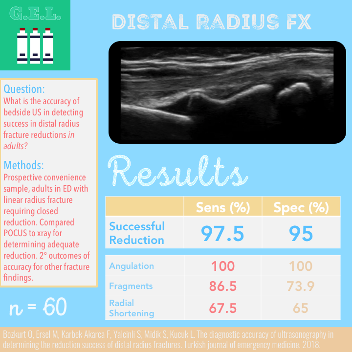
Primary Outcome – Test characteristics for determining successful reduction
Sensitivity 97.5% (CI 86.8 – 99.9%)
Specificity 95% (CI 75.1 – 99.9%)
Secondary Outcomes
US for determining direction of angulation
- Sensitivity 100% (CI 93.3 – 100%)
- Specificity 100% (CI 59.4 – 100%)
US for determining presence of fragments
- Sensitivity 86.5% (CI 71.2 – 95.5%)
- Specificity 73.9% (CI 51.6 – 89.8%)
US for determining radial shortening
- Sensitivity 67.5% (CI 50.9 – 81.4%)
- Specificity 65% (40.8 – 84.6%)
Other Findings
98% of the attempts that were thought to be successful on ultrasound were confirmed successful by xray.
Ultrasound had 1 false positive – US showed successful reduction, but xray did not. Authors: “Xray probably had an artifact…due to the plaster cast”. Ultrasound also had 1 false negative. Authors: “No comment.”
Volar angulation and multiple fragment associated with reduced success of reduction (on both xray and POCUS). Radial shortening on xray associated with less success, but not for shortening seen on ultrasound. This could be due to the very low sensitivity and specificity that was demonstrated for the ultrasound assessment of radial shortening.
Limitations
Usual things: Single operator, single center in Turkey, small population
Some room for bias if EM resident performing the ultrasound was present for the reduction attempt and could have witnessed the orthopedic residents perceived effectiveness of the reduction. Conceivable, if he thought it was likely reduced, could have stopped after finding an image of aligned cortex whereas if convinced it was poorly reduced, could have continued searching until finding an area that looked malaligned.
Remember that although the EM resident chose which images to capture, these were ultimately interpreted by a physician not involved in the acquisition. This is contrary to typical POCUS where you integrate the POCUS information into your clinical care for the patient.
Single physicians both doing the ultrasounds, interpreting the ultrasounds, and interpreting the xray. Would have been better to at least have multiple interpreters and to calculate a interrater reliability.
These patients already had an xray performed for diagnosis of the fracture. Future studies might look to see if ultrasound can be used to diagnosis, then guide reduction.
Did not attempt to measure the degree of angulation, which may be important for operative planning.
Discussion
Replacement versus supplementation. I don’t think that ultrasound can replace the xray for distal forearm fractures. Surgeons are dependent on them for good reason, you can get a lot more information. Remember that ultrasound can only assess the closest piece of bone – once the sound wave are attenuated there, you won’t be able to see underneath it. Although theoretically possible, it is difficult to determine some important characteristics of the fracture on ultrasound, merely because the sound waves cannot get past the bone cortex. Angulation, shortening, intraarticular fractures – these are all characteristics that contribute to a determination as to whether operative management may be necessary. Therefore, you are probably going to need to get an xray at some point. Where I think ultrasound can help is if you do not have fluoroscopy. This can serve as a great bedside tool to assess your reduction prior to obtaining the post-reduction xray films. It may take some practice before you get good at this. It’s easy enough to start checking with ultrasound before you get your xray or use fluoro and then compare them. Then you will be ready if you ever don’t have those tools!
Best study would be one in which patients were randomized to two groups – in intervention group they were allowed to use ultrasound in addition to xray, with outcomes being reduction time, repeat attempts, additional xrays.
Take Home Points
1. Point of care ultrasound appears to accurately determine success of reduction of distal radius fractures in adults.
2. Xray offers additional valuable information and therefore will likely still be necessary in most situations.
More Great FOAMed on this Topic
Academic Emergency Medicine Early Access Podcast – POCUS in Pediatric Forearm Fractures
Emergency Medicine Cases – POCUS for Distal Radius Fractures
Ultrasound Podcast – Ultrasound of Radius Fracture! What?
Our score

Expert Reviewer for this Post

Christopher D Thom, MD RDMS @ThomCt9k
Assistant Professor of Emergency Medicine and Assistant Director of Emergency Ultrasound at the University of Virginia
Cite this post as
Michael Prats, MD. POCUS in the Reduction of Distal Radius Fracture. Ultrasound G.E.L. Podcast Blog. Published on April 15, 2019. Accessed on February 07, 2020. Available at https://www.ultrasoundgel.org/66.


