Author: Joshua Bloom, MD, PhD // Reviewed by: Anthony DeVivo, DO (@anthony_devivo, EM-Critical Care Fellow, Icahn School of Medicine- Mount Sinai Hospital), Alex Koyfman, MD (@EMHighAK), Brit Long, MD (@long_brit) and Manpreet Singh, MD (@MprizzleER)
Welcome back to Unlocking Common ED Procedures! Today, we focus on arterial lines in the ED.
Check out our new downloadable procedure card with QR code link to the article. Print them out and be ready to go over it with your learners!
Case
An 85-year-old male presents to your emergency department (ED) in respiratory distress, hypothermic, tachypneic, hypoxic, and with diffuse rales on lung exam. He is full code and consents to invasive interventions. He has severe heart failure with reduced ejection fraction (EF 5%), and your clinical evaluation in conjunction with early testing suggest mixed cardiogenic and distributive shock secondary to sepsis. The patient is hemodynamically resuscitated, intubated for hypoxic respiratory failure, and started on two vasopressor medications (norepinephrine and dobutamine) with a goal mean arterial pressure (MAP) of 65. As he is evaluated for admission to the intensive care unit, his non-invasive blood pressure readings become erratic, with the following readings from the past 12 minutes (cycled every 3 minutes). Note that no medication titrations were performed during this time period.
23:45- 95/52 (MAP 66)
23:48- 75/48 (MAP 57)
23:51- –/– (MAP –)
23:54- 93/58 (MAP 70)
23:57- 66/46 (MAP 53)
You reassess the patient and find a bradycardic, irregular pulse in the right femoral artery. You are concerned the patient is becoming hemodynamically unstable and needs titration of his vasopressor medications; however, your noninvasive measurements appear unreliable and inconsistent. What is your next step?
Background
Accurate measurement of blood pressure is vital, particularly during the management of a critically ill patient. Significant abnormalities of blood pressure–especially hypotension–can herald organ ischemia and end-organ dysfunction and is frequently managed with the use of vasoactive medications. There are numerous methods available for blood pressure measurement, and three that you might be familiar with in the ED. Each of these methods has advantages and disadvantages1,2.
- An auscultatory method, where a sphygmomanometer is pressurized and depressurized while auscultating a distal vascular bundle for Korotkoff sounds to determine systolic and diastolic pressure, frequently at the brachial artery.
- An automated oscillatory method, where a detector within a depressurizing cuff. recognizes oscillations in vascular bundles and derives a systolic/diastolic and mean arterial pressure reading.
- Invasive blood pressure monitoring using an intra-arterial catheter and transducer3.
For lower acuity patients and initial triage vital signs in the emergency department, non-invasive blood pressure monitoring (auscultation or oscillometry) is usually sufficient to guide initial medical management. However, since these methods are reliant on intermittent measurements of distal circulation, they can prove insufficient in patients with circulatory compromise or with dynamic alterations in blood pressure. As seen in the case above, a patient in severe shock with poor distal circulation can have inconsistent or even unreadable non-invasive blood pressure readings, and in the setting of decompensated shock dynamic blood pressure medication adjustments may be needed.
This article will focus on up-to-date evidence-based indications, contraindications, methods, and tips for placing arterial lines in the ED.
Why Arterial Lines?
Arterial lines are commonly used in the ICU and OR, where patients often require close monitoring. However, adoption of this technique in the ED is far from universal. A landmark retrospective study by Lehman and colleagues in the Beth Israel Deaconess Medical Center comparing over 27,000 simultaneously obtained blood pressure measurements in ICU patients indicated that non-invasive systolic pressure readings were inaccurate in hypotensive patients compared to invasive measurements, but MAPs (arguably the measurement of choice) were comparable1. An observational study has even suggested lower extremity non-invasive blood pressure readings may produce a reliable MAP4. Smaller studies have produced similar findings in pediatric surgical patients and adult cardiac patients5,6. However, some studies have indicated that non-invasive MAP can be inaccurate compared to invasive measurements in optimally damped arterial lines7 or in patients on vasoactive medications8. It is also important to remember that systolic and diastolic pressures are important measures in multiple critical conditions (e.g. hemorrhagic stroke), and non-invasive measurements may be insufficient to titrate vasoactive medications in these patients. Critical patients often present to the ED before admission to the ICU or OR, and studies have suggested that there is no increased harm in initiating invasive blood pressure monitoring as part of ED management9. The Society for Academic Emergency Medicine has issued recommendations for arterial line placement in the emergency department and suggested that practitioners be familiar with these techniques10.
Indications
- Continuous blood pressure monitoring: invasive arterial blood pressure monitoring is often considered the gold standard for blood pressure measurement and gives second-to-second measurements allowing for close titration of blood pressure with vasoactive medications.
- Hemodynamic monitoring: invasive blood pressure waveforms have been used as surrogate measures of hemodynamic status. For example, significant respiratory variation can correspond to hypovolemia and fluid responsiveness.
- Arterial blood sampling: an arterial line can be used for recurrent blood sampling, especially if serial arterial blood gases or other tests are required. Given the potential for complications compared to arterial or venous phlebotomy, an arterial line should rarely be placed for sampling alone. It should be noted that many of the principles, risks, and contraindications discussed later in the article also apply to arterial phlebotomy.
Contraindications
There are some general contraindications to arterial line placement, as follows11.
- Vascular compromise at or distal to the desired site. Arterial catheters can occlude 100% of the vascular lumen in small-bore arteries12, and collateral circulation is needed to maintain perfusion of distal tissues. The modified Allen test has been used to examine collateral circulation distal to the radial artery, although it has limitations13,14. A corresponding test exists for arterial supply of the foot, which also often has significant collaterals15. If collateral circulation is impaired, another site should be chosen for arterial catheterization.
- Vascular abnormality at the desired site. This can include intra-arterial structures (e.g. thrombus, stent), vascular wall abnormalities (eg. atherosclerosis), anatomic abnormalities (e.g. surgical modification, fistula, or arterio-venous malformation), or systemic vascular diseases (eg. vasospasm secondary to the Reynaud phenomenon). Cannulation in these cases can risk distal tissue ischemia.
- Infection in overlying tissues. Although the risk of infection from arterial cannulation is much lower than venous cannulation, arterial lines should not be placed through cellulitis or an abscess, or with known surrounding soft tissue infection.
- Coagulopathy (relative contraindication). In the coagulopathic patient, arterial bleeding can occur and could be difficult to control. Avoid placing an arterial line in the following patients:
- INR ≥ 3
- aPTT ≥100 sec
- Platelets <50 x109/L
- Recent administration of thrombolytics
- Strongly consider the need for an arterial line in therapeutically anticoagulated patients
Considerations
- Use local anesthesia even in unconscious patients. There is evidence that it decreases vasospasm, and local infiltration should not significantly obscure the vessel, especially under dynamic ultrasound guidance16.
- Dynamic ultrasound guidance has proven superior to palpation for radial artery cannulation, with higher first-time success rates and lower failure rates. Use dynamic ultrasound guidance whenever possible17,18.
- Patients may have tough, thick, or scarred skin overlying an otherwise desirable insertion site. A dermatotomy (small skin incision) can be performed in these cases to decrease the risk of skin plugging or occlusion of the catheter. This can be performed after administration of local anesthesia.
- Site-specific considerations:
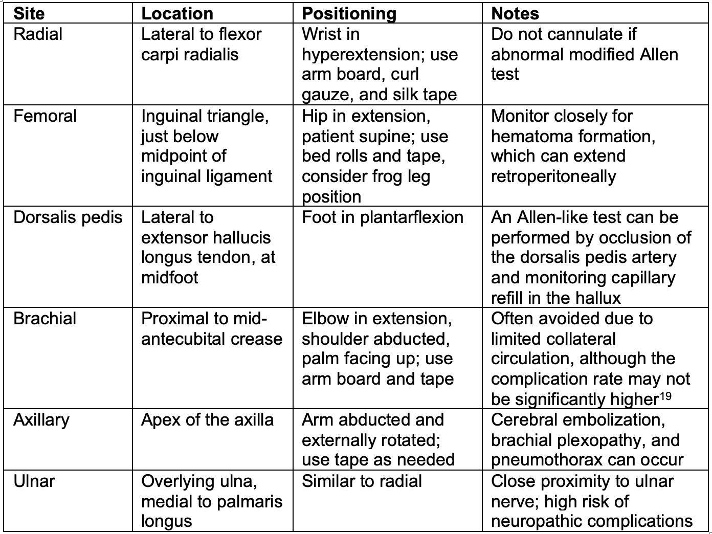
The Procedure
Please note that the goal of this procedure outline is to familiarize you with the general principles of arterial lines and their applications. Your hospital’s apparatus and guidelines may vary, and you should follow best practice guidelines at your institution.
- Materials needed
- Antiseptic site prep (chlorhexidine or iodine)
- Sterile gloves
- Sterile towels or drape
- Depending on site, items to assist with patient positioning
- Radial/brachial: arm board, silk tape, curl gauze or ace bandage
- Femoral: Bed rolls or blankets, silk tape
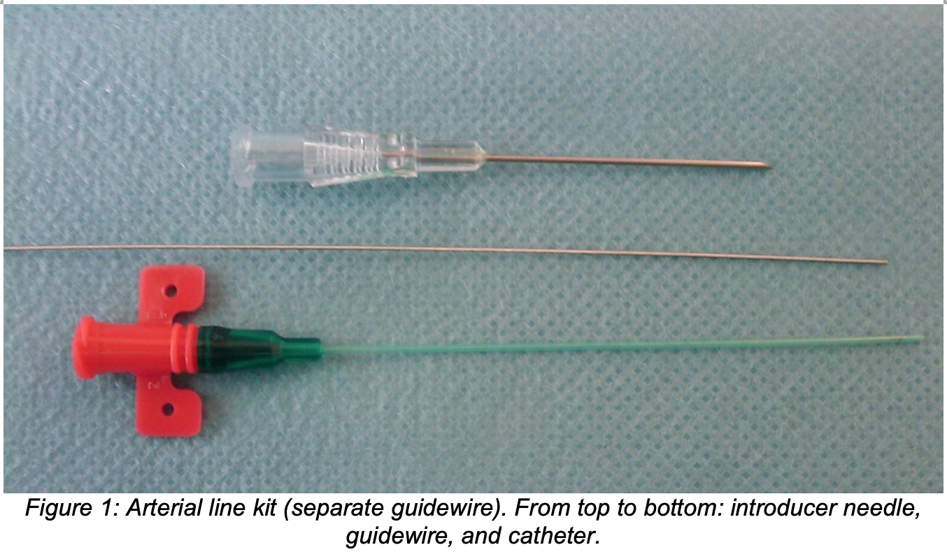
- Arterial line insertion kit. Generally, it will include an angiocatheter-style setup with a catheter installed over a needle, and either an integral or separate guidewire. A direct puncture approach can also be used, which does not require the guidewire.
-
- 1% lidocaine without epinephrine, subcutaneous needle, syringe
- Transducer, arterial line non-compliant tube set, 500mL sterile saline with pressure bag, compatible monitor and wires; make sure the tubing is flushed prior to starting the procedure, as described below
- Ultrasound (linear probe) with sterile probe cover
- Non-absorbable suture (2-0 or 3-0 silk/nylon) and transparent dressing for securing the catheter
- General procedure20
- Position patient and locate anatomy by palpation. If feasible, use ultrasound to confirm location, depth, and path of the desired artery, and make sure your desired insertion point is free of calcifications, thrombus, pseudoaneurysm, or an arteriovenous malformation.
- Don mask with eye shield, sterile gown, and gloves; assistants should also wear masks with eye shields in case of brisk arterial bleeds.
- Sterilize site with antiseptic prep solution. Be sure to sterilize broadly to allow for palpation, small adjustments, and dynamic ultrasound guidance.
- Anesthetize the skin and subcutaneous space superficial to the artery by local infiltration with 1% lidocaine. Draw back prior to injection to ensure your injection is not intravascular.
- Insert the arterial catheter under dynamic ultrasound guidance (or palpation if ultrasound is unavailable). We advise using a 30-45º angle for all first attempts.
- Direct puncture: insert an angiocatheter until pulsatile blood return is seen. Advance the catheter until hubbed and retract/remove the needle.
- Separate guidewire: insert the needle-catheter setup until pulsatile blood return is seen. Advance the catheter into the vessel slightly until blood return ceases. Remove the needle. If pulsatile blood return does not resume, retract the catheter slightly until it does. Once it does, advance the separate guidewire far past the tip of the catheter into the vessel, and advance the catheter until hubbed. Remove the guidewire.
- Integral guidewire: insert the needle-catheter-guidewire setup until pulsatile blood return is seen. Decrease the angle of insertion without advancing the catheter. If blood return is lost, advance the needle-catheter-guidewire setup slightly until blood return is seen again. Using the tab, advance the guidewire into the artery. Advance the catheter until hubbed, and remove the needle-guidewire components.
- After removal of the needle/guidewire, hold pressure proximally to avoid brisk arterial blood return.
- Connect pre-flushed arterial tubing and transducer to the arterial catheter. Have an assistant connect the transducer setup to the monitor.
- Secure the catheter with sutures (figure of 8 or horizontal mattress technique, anchored to skin) and a transparent dressing.
- Transducer
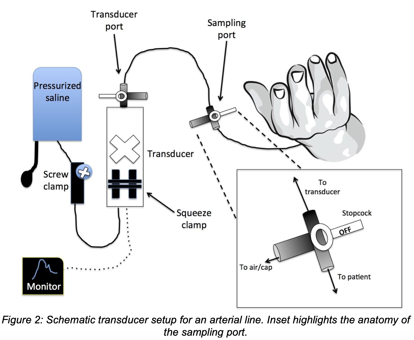
The transducer and tubing should be set up and flushed with sterile saline prior to arterial cannulation. Since the transducer apparatus will be connected to an intravascular line, it should be prepared aseptically. Remember, any stopcock on the tubing set will CLOSE the apparatus in the direction it is pointing.
-
- Spike your saline bag with the arterial tubing and release any clamps, including the pre-transducer screw clamp. Turn the transducer port stopcock OFF toward the patient side. Holding the bag upside down, evacuate air from the bag by holding the squeeze clamp open.
- Pressurize your bag of saline by inflating the pressure cuff to ~300mmHg. Lock the pressure by closing the stopcock/clamp toward the pressure bag.
- Evacuate air and bubbles from the tubing by holding the squeeze clamp. Utilize the stopcocks to ensure air and bubbles are evacuated from both the pressure bag side and the patient side of the transducer. Cap any open ports of tubing to ensure a closed system.
- At this point the tubing can be attached to the catheter in a cannulated patient.
- Position the transducer at or around the phlebostatic axis (4th intercostal, midaxillary) and secure with tape or a transducer clip assembly.
- You need to zero the transducer to atmospheric pressure. Turn the stopcock of the transducer port OFF to the patient and uncap the attached port. Holding the squeeze clamp should cause pressurized saline to squirt from the port. Connect the monitor wires and zero the line on the monitor.
- Turn the stopcock OFF to the port and cap the port. The monitor should begin showing an arterial waveform.
- Arterial blood sampling
- Turn the sampling port stopcock OFF to the transducer. Note that continuous pressure monitoring will not be possible during sampling.
- Uncap the sampling port and clean with chlorhexidine solution. Using a vacutainer or syringe, evacuate/waste 10-12 mL liquid from the patient side of the line to avoid contamination of blood samples with saline. If evacuating with a syringe, close the port and line by leaving the stopcock at 45º between port and transducer while switching syringes.
- Using a vacutainer or heparinized syringe, withdraw arterial blood for testing.
- After you are finished, flush the patient side of the sampling port with saline, and turn the stopcock OFF to the port. Clean and recap the port.
- Arterial line removal
Although not always applicable during the timeline of emergency care, catheters that are deemed no longer necessary or those that develop a complication should be removed immediately.- Flush the catheter with saline or draw arterial blood back into the catheter.
- Cut/remove any suture or tape securing the catheter.
- Clean the site with chlorhexidine.
- While applying proximal and local pressure with sterile 4×4 gauze, slowly remove the catheter. Inspect it quickly for any defects that may suggest a particulate embolism.
- Hold pressure at the site for 5-10 minutes. In a coagulopathic patient, the pressure may need to be held for double the time or more, and the patient should be examined serially within the first 24 hours for hematoma formation or bleeding.
Post-Procedural Complications21,22
- Arterial vasospasm
Arteries are made of smooth muscle that can constrict or dilate in response to a variety of stimuli. Local trauma tends to prompt vasoconstriction, which is adaptive in case of traumatic arterial injury. Unfortunately, this effect can also significantly increase the discomfort associated with arterial line placement and can cause procedural failure23. Arterial vasospasm is also associated with thrombotic events after both failed and successful catheterizations. Avoid this complication by avoiding multiple attempts on the same artery, using a catheter appropriately sized to your artery, and aborting the procedure early if distal pulses or waveforms are lost. Using local anesthesia can also decrease the incidence of this complication. - Thrombosis
Placement of an intraarterial catheter involves vascular injury, intravascular foreign body disrupting flow, and local vascular stasis. Thrombosis can easily occur under these conditions. Arterial thrombosis is a potentially limb-threatening complication and can cause distal ischemia, especially if the patient is vasculopathic or the limb lacks good collaterals. You can reduce this complication in your ED arterial line placements by avoiding vasospasm during placement and using an appropriate catheter to artery ratio (smallest catheter, largest diameter of artery). - Embolism
Multiple types of emboli are possible during arterial cannulation. Thrombi that form around a catheter or insertion site can embolize distally and cause limb ischemia. Damaged catheters can fragment, causing particulate emboli that can have similar effects. Air introduced during the procedure can also embolize and cause distal ischemia and has even been reported to embolize proximally and cause ischemia in other sites such as cerebral circulation (stroke)24. Note that embolization can occur during introduction of the catheter, while the catheter is in place, or during removal of the catheter. Air emboli can also be introduced via the transducer set or during blood sampling. Avoid this complication by routine examination of circulation in the cannulated limb, evacuating air from all parts of the catheter and transducer setup, and inspecting catheters for damage before insertion and after removal. - Intra-arterial infusion
Medications are not routinely administered into an arterial catheter. Intra-arterial medication administration can be benign or devastating depending on the medication administered25. For example, administration of a vasoconstricting medication, cytotoxic medication, or a poorly soluble medication that precipitates can cause distal limb ischemia. Any observed administration of intra-arterial medication should be aborted immediately, and you should have a low threshold to consult appropriate specialists (vascular surgery, interventional radiology, toxicology) if you believe the administered medication can be limb threatening. - Structural complications
Trauma to an artery wall can have immediate and long-term structural complications. Dissection has been reported, which can result in vascular compromise and limb ischemia26. Pseudoaneurysms and arteriovenous fistulae have also been observed27. These are usually uncomfortable for the patient, may complicate future vascular procedures in this area, and can acutely cause hematoma formation and compartment syndrome. Avoid structural complications by using appropriately sized catheters and avoiding multiple attempts on the same artery. Using dynamic ultrasound guidance can reveal any pre-existing structural abnormalities before the procedure; if you observe this, you should select a new site for cannulation. - Infection
Conventional wisdom has held that arterial catheters are at lower risk for bloodstream infection compared to venous catheters. However, arterial lines are still an important source of catheter-related bloodstream infections28. Avoid this complication by using strict sterile technique during catheter insertion, and aseptic technique should be used while setting up and changing the transducer. Radial cannulation seems to have a decreased risk of bloodstream infection compared to femoral sites. - Bleeding
Arterial cannulation inevitably causes bleeding, and some bleeding is anticipated during the procedure. Avoid severe iatrogenic blood loss by holding good pressure during catheterization and making sure all your equipment is readily available before you start. Be aware that unnecessary iatrogenic blood loss can also occur during blood sampling; if possible, sampling from the port closest to the patient can mediate this29. The insertion site should be examined serially after the procedure, as arterial rupture and hematoma formation can occur and potentially cause a compartment syndrome, severe blood loss, or limb ischemia.
The Arterial Waveform
- You should feel comfortable with the basic principles of an arterial waveform. The elements of an arterial waveform are summarized below.
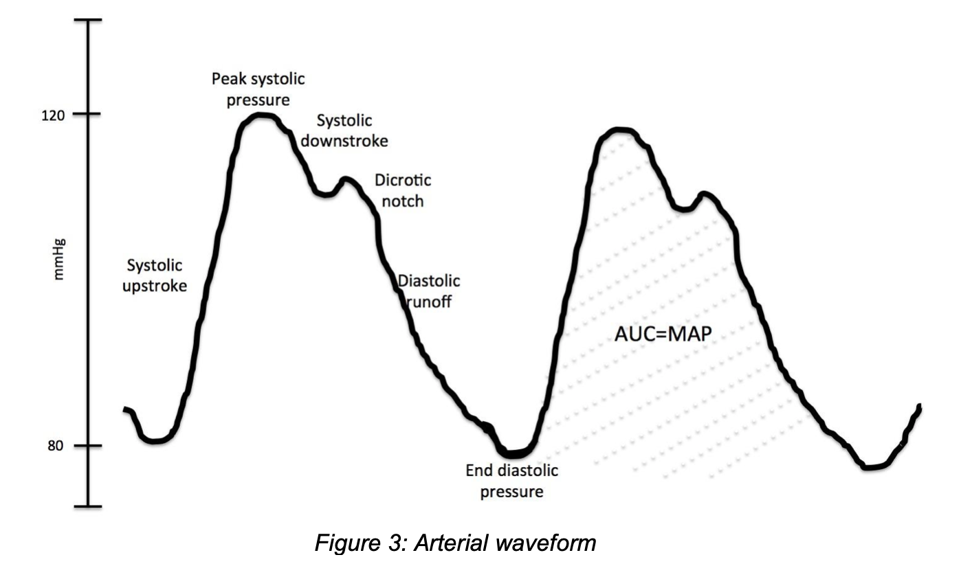
- The dicrotic notch, or incisura, is caused by the closure of the aortic valve.
- Most arterial line monitors will average the pressures and MAP over several waveforms to produce the reported value.
- The arterial waveform is determined by cardiac output and vascular resistance. One study of cardiac bypass patients indicated peripheral cannulation sites (e.g. radial, dorsalis pedis) exhibit steeper systolic upstrokes, higher pulse pressures, and higher systolic pressures, whereas diastolic and mean arterial pressure measurements seem unaffected30. A more recent study in patients on high-dose norepinephrine had distal arterial monitoring underestimate multiple blood pressure measures, including MAP31.
- You may observe respiratory variations in the pressure waveforms. A large variation can suggest fluid responsiveness in hypovolemic patients. There are devices available that measure indices such as systolic pressure variation (SPV) and pulse pressure variation (PPV)32,33.
- All components of the transducer apparatus produce a dampening effect on the arterial blood pressure waveform34,35. Appropriate dampening produces a square waveform after a rapid flush of the non-compliant tubing with saline, with 1-3 “ringing” oscillations. An over-dampened waveform appears artificially flattened with a decreased pulse pressure and loss of ringing after a rapid flush and can be caused by bubbles or kinks in the tubing. An under-dampened waveform has an exaggerated pulse pressure with excessive ringing and can be caused by excessive tubing length with multiple stopcocks.
Case Review
This patient could certainly benefit from an arterial line. As you prepare for the invasive procedure, though, you instruct a colleague to troubleshoot the non-invasive blood pressure measurement device and equipment. They exchange the patient’s cuff for an appropriately-sized one and make sure it is appropriately positioned. A few minutes later you insert a left radial arterial line aseptically under dynamic ultrasound guidance without complication. The patient’s invasive blood pressure readings show a consistent MAP in the 50’s, which is below your goal for this patient. You increase the patient’s dobutamine drip, which stabilizes the MAP to 65-70. Your patient is transported to the cardiac ICU without incident.
Rapid Procedure Review
- Position patient and site of interest.
- Prepare your equipment, including a fully connected transducer setup that has been prepared aseptically, flushed, and evacuated of air.
- Palpate the pulse of the artery you intend to cannulate; examine distal collateral circulation (e.g. modified Allen’s test) as needed. Use ultrasound to confirm the location and integrity of your artery.
- Don sterile gown and gloves, apply sterile drape, and sterilize site with antiseptic.
- Anesthetize the site with local anesthetic.
- Insert the arterial catheter under dynamic ultrasound guidance. Remove any needle or guidewire and hold pressure to avoid brisk arterial bleed.
- Connect the transducer to the catheter and have an assistant connect to the monitor.
- Suture the catheter in place and secure with a transparent dressing.
References/Further Reading:
- Lehman, L.-W. H., Saeed, M., Talmor, D., Mark, R. & Malhotra, A. Methods of blood pressure measurement in the ICU. Crit. Care Med. 41, 34–40 (2013).
- Magder, S. A. The highs and lows of blood pressure: toward meaningful clinical targets in patients with shock. Crit. Care Med. 42, 1241–1251 (2014).
- Rutten, A. J., Ilsley, A. H., Skowronski, G. A. & Runciman, W. B. A comparative study of the measurement of mean arterial blood pressure using automatic oscillometers, arterial cannulation and auscultation. Anaesth Intensive Care 14, 58–65 (1986).
- Lakhal, K., Macq, C., Ehrmann, S., Boulain, T. & Capdevila, X. Noninvasive monitoring of blood pressure in the critically ill: reliability according to the cuff site (arm, thigh, or ankle). Crit. Care Med. 40, 1207–1213 (2012).
- Meidert, A. S. et al. Accuracy of oscillometric noninvasive blood pressure compared with intra-arterial blood pressure in infants and small children during neurosurgical procedures: An observational study. Eur J Anaesthesiol 36, 400–405 (2019).
- Seidlerová, J., Tůmová, P., Rokyta, R. & Hromadka, M. Factors influencing the accuracy of non-invasive blood pressure measurements in patients admitted for cardiogenic shock. BMC Cardiovasc Disord 19, 150–10 (2019).
- Joffe, R., Duff, J., Garcia Guerra, G., Pugh, J. & Joffe, A. R. The accuracy of blood pressure measured by arterial line and non-invasive cuff in critically ill children. Crit Care 20, 177–9 (2016).
- Saherwala, A. A. et al. Correlation of Noninvasive Blood Pressure and Invasive Intra-arterial Blood Pressure in Patients Treated with Vasoactive Medications in a Neurocritical Care Unit. Neurocrit Care 28, 265–272 (2018).
- Saladino, R., Bachman, D. & Fleisher, G. Arterial access in the pediatric emergency department. YMEM 19, 382–385 (1990).
- Al-Joburi, D. W. & Farabaugh, E. A. Vascular Access. Clerkship Directors in Emergency Medicine. https://www.saem.org/cdem/education/online-education/m3-curriculum/group-emergency-department-procedures/vascular-access. Accessed November 6th, 2019.
- Scheer, B., Perel, A. & Pfeiffer, U. J. Clinical review: complications and risk factors of peripheral arterial catheters used for haemodynamic monitoring in anaesthesia and intensive care medicine. Crit Care 6, 199–204 (2002).
- Slogoff, S., Keats, A. S. & Arlund, C. On the safety of radial artery cannulation. Anesthesiology 59, 42–47 (1983).
- Allen, E. V. Thrombo-Angiitis Obliterans. Bull N Y Acad Med 18, 165–189 (1942).
- Kohonen, M., Teerenhovi, O., Terho, T., Laurikka, J. & Tarkka, M. Is the Allen test reliable enough? European Journal of Cardio-Thoracic Surgery 32, 902–905 (2007).
- Haddock, N. T., Garfein, E. S., Saadeh, P. B. & Levine, J. P. The lower-extremity Allen test. J Reconstr Microsurg 25, 399–403 (2009).
- Lightowler, J. V. & Elliott, M. W. Local anaesthetic infiltration prior to arterial puncture for blood gas analysis: a survey of current practice and a randomised double blind placebo controlled trial. J R Coll Physicians Lond 31, 645–646 (1997).
- White, L., Halpin, A., Turner, M. & Wallace, L. Ultrasound-guided radial artery cannulation in adult and paediatric populations: a systematic review and meta-analysis. Br J Anaesth 116, 610–617 (2016).
- Moussa Pacha, H. et al. Ultrasound-guided versus palpation-guided radial artery catheterization in adult population: A systematic review and meta-analysis of randomized controlled trials. Am. Heart J. 204, 1–8 (2018).
- Handlogten, K. S., Wilson, G. A., Clifford, L., Nuttall, G. A. & Kor, D. J. Brachial artery catheterization: an assessment of use patterns and associated complications. Anesth. Analg. 118, 288–295 (2014).
- Tegtmeyer, K., Brady, G., Lai, S., Hodo, R. & Braner, D. Videos in Clinical Medicine. Placement of an arterial line. The New England journal of medicine 354, e13 (2006).
- Downs, J. B., Rackstein, A. D., Klein, E. F. & Hawkins, I. F. Hazards of radial-artery catheterization. Anesthesiology 38, 283–286 (1973).
- Frezza, E. E. & Mezghebe, H. Indications and complications of arterial catheter use in surgical or medical intensive care units: analysis of 4932 patients. Am Surg 64, 127–131 (1998).
- Kristić, I. & Lukenda, J. Radial artery spasm during transradial coronary procedures. J Invasive Cardiol 23, 527–531 (2011).
- Chang, C. et al. Air embolism and the radial arterial line. Crit. Care Med. 16, 141–143 (1988).
- Andreev, A., Kavrakov, T., Petkov, D. & Penkov, P. Severe acute hand ischemia following an accidental intraarterial drug injection, successfully treated with thrombolysis and intraarterial Iloprost infusion. Case report. Angiology 46, 963–967 (1995).
- Weinberg, L., Abu-Ssaydeh, D., Spanger, M., Lu, P. & Li, M. H. G. Case report: Iatrogenic brachial artery dissection with complete anterograde occlusion during elective arterial line placement. Int J Surg Case Rep 42, 269–273 (2018).
- Tosti, R., Özkan, S., Schainfeld, R. M. & Eberlin, K. R. Radial Artery Pseudoaneurysm. J Hand Surg Am 42, 295.e1–295.e6 (2017).
- O’Horo, J. C., Maki, D. G., Krupp, A. E. & Safdar, N. Arterial catheters as a source of bloodstream infection: a systematic review and meta-analysis. Crit. Care Med. 42, 1334–1339 (2014).
- Silver, M. J., Jubran, H., Stein, S., McSweeney, T. & Jubran, F. Evaluation of a new blood-conserving arterial line system for patients in intensive care units. Crit. Care Med. 21, 507–511 (1993).
- Pauca, A. L., Wallenhaupt, S. L., Kon, N. D. & Tucker, W. Y. Does radial artery pressure accurately reflect aortic pressure? Chest 102, 1193–1198 (1992).
- Kim, W. Y. et al. Radial to femoral arterial blood pressure differences in septic shock patients receiving high-dose norepinephrine therapy. Shock 40, 527–531 (2013).
- Thiele, R. H., Colquhoun, D. A., Blum, F. E. & Durieux, M. E. The Ability of Anesthesia Providers to Visually Estimate Systolic Pressure Variability Using the ‘Eyeball’ Technique. Anesth. Analg. 115, 176–181 (2012).
- Jozwiak, M., Monnet, X. & Teboul, J.-L. Pressure Waveform Analysis. Anesth. Analg. 126, 1930–1933 (2018).
- Boutros, A. & Albert, S. Effect of the dynamic response of transducer-tubing system on accuracy of direct blood pressure measurement in patients. Crit. Care Med. 11, 124–127 (1983).
- Romagnoli, S. et al. Accuracy of invasive arterial pressure monitoring in cardiovascular patients: an observational study. Crit Care 18, 575–11 (2014).
