Authors: Kamoga Dickson, MD (EM Resident Physician, Makerere College of Health Sciences, Kampala, Uganda); Jessica Pelletier, DO, MHPE (APD/Assistant Professor of EM/Attending Physician, University of Missouri-Columbia) // Reviewed by: Alex Koyfman, MD (@EMHighAK); Brit Long, MD (@long_brit)
Case
A 42-year-old male from a rural area in northeastern Uganda presents to the tertiary care emergency department with severe itching, skin changes on his legs, and episodes of blurred vision, describing “something moving” in his eye. He was referred from a primary care clinic due to worsening skin thickening and nodules over several months for which he received several antibiotics and antihistamines without symptomatic improvement. Examination reveals hyperpigmented, thickened skin with nodules. There is conjunctival injection with a partially opacified cornea in the right eye. Non-tender inguinal lymph nodes are noted. Skin biopsy ultimately confirms infection with Onchocerca (O.) volvulus, leading to a diagnosis of onchocerciasis with ocular involvement. Treatment with ivermectin is initiated, and the patient is urgently referred to ophthalmology for further treatment.
Introduction
Onchocerciasis, also known as river blindness, is a chronic infection caused by the helminthic worm O. volvulus. It is the second most common cause of infectious blindness after trachoma in the world and is transmitted through the repeated bites of the blackfly (Simulium damnosum), which acts as the vector. O. volvulus is a ubiquitous filarial worm that multiplies in freshwater bodies and solely infects humans.1 O. volvulus infection is one of the major Neglected Tropical Diseases (NTDs) that affect people living in underdeveloped countries.2 125 million people around the globe are at risk for infection, and 18 million people are infected with O. volvulus – 99% of whom reside in sub-Saharan Africa.3
Onchocerciasis is a clinical syndrome characterized by ophthalmologic (commonest), dermatologic, lymphatic, and (rarely) systemic manifestations. Clinical manifestations occur as a result of dead and dying microfilariae inciting an immunologic/inflammatory response, ultimately resulting in tissue damage and scarring. However, recent progress in onchocerciasis research has led to an improved understanding of the immunopathology of O. volvulus as well as improvements in the diagnosis and treatment of this morbid disease.
This article aims to create emergency clinician awareness of onchocerciasis as a potential cause of visual and dermatologic complaints and will highlight appropriate diagnosis and management strategies. Vision loss carries a high degree of morbidity and is associated with higher all-cause mortality,4 and it is our role as emergency providers to recognize potentially reversible causes of such adverse outcomes. Knowledge of NTDs such as onchocerciasis is essential for building an appropriate differential diagnosis in the emergency department given the rising numbers of refugees and immigrants who are moving to the United States (US) from sub-Saharan Africa (the US is the most common destination for sub-Saharan African immigrants who leave the African continent).5
Epidemiology
Onchocerciasis is endemic in 31 African countries but is also problematic in Brazil, Venezuela, and Yemen.6 By 2030, it is estimated that Nigeria followed by the Democratic Republic of the Congo and South Sudan will have the greatest disease burden.7Approximately 14.6 million infected people suffer from skin disease and 1.15 million from partial or complete blindness.6 Worldwide ongoing mass drug administration (MDA) with ivermectin, a public health measure made possible via mass donations from Merck, significantly reduces the burden of infection and associated ocular disease.8 MDA requires 12-15 years of annual treatment in endemic areas to successfully eliminate transmission and has been successful in Columbia, Ecuador, Mexico, and Guatemala.6
Risk Factors
The following are known risk factors for developing onchocerciasis:
- Male gender
- Distance less than 2 km from a riverbank
- Noncompliance with ivermectin therapy
- Age > 35 years9
It should be noted that short-term travelers to endemic areas are at low risk for contracting onchocerciasis since the development of the disease requires repeated blackfly bites.1
Life Cycle and Pathophysiology
Life Cycle
Humans are the only hosts for O. volvulus.1 The parasite has a 5-stage life cycle in which the blackfly acts as an obligate intermediate host. Infection occurs when a blackfly introduces an O. volvulus stage 3 larvae into human skin during a blood meal. The larvae reside in the skin (inside of a fibrous capsule or nodule) until reaching adulthood. The worms can live as long as 15 years, and female worms may produce microfilariae (early-stage larvae) for up to 9 of those years.10 During adulthood, the female worm sheds hundreds of thousands of microfilariae that migrate through the skin of the human host, with particular affinity for the eyes.11 The inflammatory response against dying microfilariae over years of repeated infection causes the gradual and eventually blinding sclerosal opacification of the anterior eye by local inflammation and of the posterior eye by autoimmune mechanisms.12
The O. volvulus life cycle continues on the uptake of microfilariae by the blackfly during a blood meal from a human. The microfilariae penetrate the fly’s gut and migrate to the thoracic flight muscles, where they develop into third-stage larvae and then find their way to the blackfly’s feeding apparatus. They then enter another human host during a blood meal, thus completing the cycle. Microfilariae persist in the human host for 3– 5 years, a much shorter period of time than the adult female worm.13
Figure 1. O. volvulus life cycle. Open-access image reference: By Giovanni Maki, derived from a CDC image at http://www.dpd.cdc.gov/dpdx/HTML/Filariasis.htm – Basáñez M-G, Pion SDS, Churcher TS, Breitling LP, Little MP, et al. (2006) River Blindness: A Success Story under Threat? PLoS Med 3(9): e371. doi:10.1371/journal.pmed.0030371 (image link), CC BY 2.5, https://commons.wikimedia.org/w/index.php?curid=1357738

Pathophysiologic mechanisms
Dermatological
In early stages, microfilariae enter the papillary dermis leading to inflammatory changes. Dying larvae lead to the formation of microabscess and granulomae within the dermis. Onchocercal nodules (i.e. onchocercoma) occur as a result of O. volvulus worms becoming embedded within inflammatory granulomas. Advanced stages of onchocerciasis typically manifest with acanthosis, parakeratosis, tumor melanosis, and pigment incontinence.14 Skin changes from onchocerciasis can be quite deforming and are permanent.15
Ocular
Ocular pathology involves both anterior and posterior segments, and the mechanisms are not completely understood. Dead or dying microfilariae are believed to trigger an inflammatory cascade that leads to tissue damage associated with the disease. Specifically, it is known that the sclerosing keratitis that leads to corneal blindness is mediated through modification of ICAM-1 expression and production of IL-4 and IL-13.16 Pathological complications that occur following this inflammatory process include uveitis with the formation of synechiae, secondary cataract, and glaucoma, which may be of open-angle or closed-angle forms.17 Posterior segment pathologies including chorioretinal and optic nerve damage are believed to be mediated by autoimmune processes or the persistence of microfilariae or their products in the posterior segment.18,19 Though early visual symptoms are potentially reversible, blindness from onchocerciasis is permanent.6
Clinical Presentation
Onchocerciasis causes ocular and dermatologic manifestations.
Dermatological manifestations
Normally present years before ocular manifestations arise. Manifestations include:20
- Recurrent severe pruritus
- Papular dermatitis
- Lichenified dermatitis
- Areas of skin depigmentation, especially around the shins, give rise to the name “leopard skin.” Skin depigmentation happens as a result of chronic rubbing of the skin in response to severe itchiness induced by an immunological response to dying or dead microfilariae
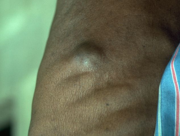
Figure 2. Onchocerciasis nodule. Onchocerciasis nodule occurring in the thorax Contributed by Prof. Gary J. Weil, MD. Washington University in St. Louis. This material is available under the Creative Commons license. Image source: Gyasi ME, Okonkwo ON, Tripathy K. Onchocerciasis. [Updated 2023 Aug 25]. In: StatPearls [Internet]. Treasure Island (FL): StatPearls Publishing; 2024 Jan-. Available from: https://www.ncbi.nlm.nih.gov/books/NBK559027/
Ocular manifestations
- Recurrent conjunctivitis
- Punctate keratitis
- Photophobia
- Progressive visual loss from either anterior (cornea scarring) or posterior segment pathologies (optic nerve and chorioretinal atrophies) or both.
- Snowflake keratitis – induced by reaction to dead microfilariae
- Corneal scarring
- Cataract
- Posterior segment diseases are related to the optic nerve and chorioretinal atrophy
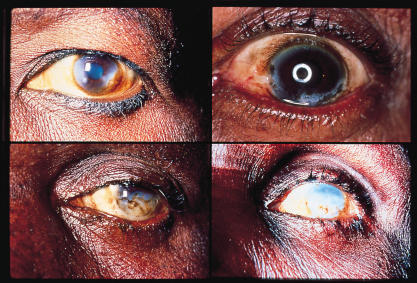
Figure 3. Sclerosing keratitis caused by onchocerciasis. Contributed by Ian Murdoch and Allen Foster. This material is available under the Creative Commons license. Image source: 1. Murdoch I, Foster A. File:Sclerosing Keratitis.Jpg.; 2001. Accessed November 17, 2024. https://eyewiki.org/File:Sclerosing_keratitis.jpg
Differential Diagnosis
- Dermatologic manifestations
- Hypersensitivity reaction
- Food allergy
- Filariasis
- Ocular manifestations
- Loa loa infection
- Vitamin A deficiency
Diagnosis
The diagnosis of onchocerciasis typically requires tissue biopsy; thus, it is unlikely that the definitive diagnosis will be made in the emergency department. Patients from endemic regions with signs and symptoms suggestive of onchocerciasis will require multidisciplinary consultation, including ophthalmology, dermatology, and infectious disease.
Table 1. Diagnostic methods that may aid in the diagnosis of onchocerciasis.
*Having the patient bend at the waist and put their head between their knees for 2 minutes prior to slit lamp examination can make it easier to visualize microfilariae in the anterior chamber.25 AC = Anterior chamber, ELISA = enzyme-linked immunosorbent assay, NS = normal saline, PCR = polymerase chain reaction
Management
Oral Ivermectin26
- 150 mcg/kg given once or twice yearly for 10 to 15 years to match the lifespan of the adult worm is the standard treatment.
- Ivermectin paralyzes or kills microfilariae but does not kill adult worms, though it temporarily reduces female worm fertility.
- It effectively decreases blindness and skin disease associated with onchocerciasis.
- Daily treatments are unnecessary, as they offer no proven benefits over periodic dosing, and higher doses may be harmful.
- More frequent dosing (every 3-6 months) may help reduce symptoms faster and sterilize the adult worm more rapidly, especially for those not returning to endemic areas.
Doxycycline26
- Doxycycline targets Wolbachia, an endosymbiotic, intracellular rickettsia-like bacteria, which is important for embryogenesis and for the survival of adult worms.
- Doxycycline does not kill the microfilaria for which another drug has to be used concurrently.
- Dosing is 100-200 mg once daily for 6 weeks.
- Ivermectin should be started at least 1 week before doxycycline for optimal benefits.
Moxidectin
- May be superior to ivermectin in reducing skin microfilariae load after 12 months of a single oral dose of 8 mg.27
- Interferes with glutamate-gated chloride ion channels, interfering with neurotransmission and leading to paralysis of the parasite.
- May actually kill and/or sterilize the adult worm.
- Long-term population-level data is still needed.21
Older treatments, including suramin and diethylcarbamazine
- Are avoided as less toxic and effective alternative treatments are available.
- Diethylcarbamazine may accelerate the development of blindness.28
Prognosis
- Often ocular and dermatologic complications of onchocerciasis improve with ivermectin treatment, especially if the disease is not advanced.
- When the disease is advanced, blindness is irreversible.
- Dermatological manifestations of advanced disease such as skin depigmentation and atrophy are irreversible.15
Prevention
Compliance with MDA is critically important, as successful campaigns require 80% of an at-risk population in an endemic area to receive bi-annual ivermectin therapy for at least 10 years.29 In addition to compliance with public health campaigns that administer ivermectin, those in endemic areas should:
- Keep windows and doors closed.
- Sleep under treated bed nets.
- Avoid contact with fresh running water if possible.
- Wear loose-fitting full-coverage clothing and use DEET-containing insect repellent when in proximity to fresh running water.
- Avoid walking barefoot.30,31
Pearls and Pitfalls
- Onchocerciasis remains endemic in many low-resource regions, with the highest burden in sub-Saharan Africa.
- Presenting symptoms usually include skin nodules, severe pruritis, dermatitis, and depigmentation followed by vision loss. Patients may or may not recall a preceding blackfly bite.
- Emergency clinicians in endemic areas or those treating patients from endemic areas should be vigilant for the disease in patients with suggestive symptoms and seek early consultation from ophthalmology, dermatology, and infectious disease.
- Ivermectin is typically the first-line therapy. Patients must understand the importance of long-term therapy (10-15 years).
- Ongoing research aims to develop more effective treatments to target adult worms and reduce treatment duration.
References
- Winthrop KL, Furtado JM, Silva JC, Resnikoff S, Lansingh VC. River blindness: an old disease on the brink of elimination and control. J Glob Infect Dis. 2011;3(2):151-155. doi:10.4103/0974-777X.81692
- USAID. Onchocerciasis.; 2024. Accessed November 16, 2024. https://www.neglecteddiseases.gov/about/usaid-targeted-diseases/onchocerciasis/
- Etya’ale D. Vision 2020: update on onchocerciasis. Community Eye Health. 2001;14(38):19-21.
- Ehrlich JR, Ramke J, Macleod D, et al. Association between vision impairment and mortality: a systematic review and meta-analysis. Lancet Glob Health. 2021;9(4):e418-e430. doi:10.1016/S2214-109X(20)30549-0
- Lorenzi J, Batalova J. Sub-Saharan African Immigrants in the United States.; 2022. Accessed November 18, 2024. https://www.migrationpolicy.org/article/sub-saharan-african-immigrants-united-states-2019#:~:text=Globally%2C%20approximately%2028.3%20million%20sub,%2C%20and%20Canada%20(435%2C000).
- World Health Organization (WHO). Onchocerciasis.; 2022. Accessed November 17, 2024. https://www.who.int/news-room/fact-sheets/detail/onchocerciasis#:~:text=Onchocerciasis%20occurs%20mainly%20in%20tropical,Uganda%2C%20United%20Republic%20of%20Tanzania.
- Vinkeles Melchers NVS, Stolk WA, Van Loon W, et al. The burden of skin disease and eye disease due to onchocerciasis in countries formerly under the African Programme for Onchocerciasis Control mandate for 1990, 2020, and 2030. Ramos AN, ed. PLoS Negl Trop Dis. 2021;15(7):e0009604. doi:10.1371/journal.pntd.0009604
- Taylor MJ, Awadzi K, Basáñez MG, et al. Onchocerciasis Control: Vision for the Future from a Ghanian perspective. Parasit Vectors. 2009;2(1):7. doi:10.1186/1756-3305-2-7
- Kifle B, Woldemichael K, Nigatu M. Prevalence of Onchocerciasis and Associated Factors among Adults Aged ≥ 15 Years in Semen Bench District, Bench Maji Zone, Southwest Ethiopia: Community Based Cross-Sectional Study. Adv Public Health. 2019;2019:1-9. doi:10.1155/2019/7276230
- Centers for Disease Control and Prevention (U.S.). Onchocerciasis.; 2024. Accessed November 17, 2024. https://www.cdc.gov/dpdx/onchocerciasis/index.html
- Little M, Breitling L, Basáñez MG, Alley E, Boatin B. Association between microfilarial load and excess mortality in onchocerciasis: an epidemiological study. The Lancet. 2004;363(9420):1514-1521. doi:10.1016/S0140-6736(04)16151-5
- Hall LR, Pearlman E. Pathogenesis of Onchocercal Keratitis (River Blindness). Clin Microbiol Rev. 1999;12(3):445-453. doi:10.1128/CMR.12.3.445
- Somorin AO. Onchocerciasis. Int J Dermatol. 1983;22(3):182-188. doi:10.1111/j.1365-4362.1983.tb03361.x
- Frallonardo L, Di Gennaro F, Panico GG, et al. Onchocerciasis: Current knowledge and future goals. Front Trop Dis. 2022;3:986884. doi:10.3389/fitd.2022.986884
- Murdoch ME, Hay RJ, Mackenzie CD, et al. A clinical classification and grading system of the cutaneous changes in onchocerciasis. Br J Dermatol. 1993;129(3):260-269. doi:10.1111/j.1365-2133.1993.tb11844.x
- Berger RB, Blackwell NM, Lass JH, Diaconu E, Pearlman E. IL-4 and IL-13 regulation of ICAM-1 expression and eosinophil recruitment in Onchocerca volvulus keratitis. Invest Ophthalmol Vis Sci. 2002;43(9):2992-2997.
- Egbert PR. Onchocerciasis: a potential risk factor for glaucoma. Br J Ophthalmol. 2005;89(7):796-798. doi:10.1136/bjo.2004.061895
- Semba RD, Murphy RP, Newland HS, Awadzi K, Greene BM, Taylor HR. Longitudinal Study of Lesions of the Posterior Segment in Onchocerciasis. Ophthalmology. 1990;97(10):1334-1341. doi:10.1016/S0161-6420(90)32413-2
- Banla M, Tchalim S, Karabou PK, et al. Sustainable Control of Onchocerciasis: Ocular Pathology in Onchocerciasis Patients Treated Annually with Ivermectin for 23 Years: A Cohort Study. Bejon P, ed. PLoS ONE. 2014;9(6):e98411. doi:10.1371/journal.pone.0098411
- Ali MM, Baraka OZ, AbdelRahman SI, et al. Immune Responses Directed against Microfilariae Correlate with Severity of Clinical Onchodermatitis and Treatment History. J Infect Dis. 2003;187(4):714-717. doi:10.1086/367709
- Babalola O. Ocular onchocerciasis: current management and future prospects. Clin Ophthalmol. Published online October 2011:1479. doi:10.2147/OPTH.S8372
- Enk CD. Onchocerciasis—river blindness. Clin Dermatol. 2006;24(3):176-180. doi:10.1016/j.clindermatol.2005.11.008
- Centers for Disease Control and Prevention (U.S.). Diagnosis of Onchocerciasis.; 2024. https://www.cdc.gov/filarial-worms/testing/index.html#:~:text=Examine%20a%20skin%20snip%E2%80%94a,examine%20it%20for%20adult%20worms.
- Mand S, Marfo-Debrekyei Y, Debrah A, et al. Frequent detection of worm movements in onchocercal nodules by ultrasonography. Filaria J. 2005;4(1):1. doi:10.1186/1475-2883-4-1
- McLeod SD. Parasitic Keratitis. In: Ophthalmology. Elsevier; 2009:274-278. doi:10.1016/B978-0-323-04332-8.00038-X
- Showler AJ, Nutman TB. Imported onchocerciasis in migrants and travelers. Curr Opin Infect Dis. 2018;31(5):393-398. doi:10.1097/QCO.0000000000000483
- Opoku NO, Bakajika DK, Kanza EM, et al. Single dose moxidectin versus ivermectin for Onchocerca volvulus infection in Ghana, Liberia, and the Democratic Republic of the Congo: a randomised, controlled, double-blind phase 3 trial. The Lancet. 2018;392(10154):1207-1216. doi:10.1016/S0140-6736(17)32844-1
- Awadzi K, Ottesen EA, Francis H. The Mazzotti Reaction Following Treatment of Onchocerciasis with Diethylcarbamazine: Clinical Severity as a Function of Infection Intensity. Am J Trop Med Hyg. 1985;34(3):529-536. doi:10.4269/ajtmh.1985.34.529
- Mutono N, Basáñez MG, James A, et al. Elimination of transmission of onchocerciasis (river blindness) with long-term ivermectin mass drug administration with or without vector control in sub-Saharan Africa: a systematic review and meta-analysis. Lancet Glob Health. 2024;12(5):e771-e782. doi:10.1016/S2214-109X(24)00043-3
- Centers for Disease Control and Prevention (U.S.). About Onchocerciasis.; 2024. Accessed November 17, 2024. https://www.cdc.gov/filarial-worms/about/onchocerciasis.html
- Abdissa D, Kebede Y, Sudhakar M, et al. Community’s knowledge, perceptions and preventive practices on Onchocerciasis in Jimma zone, Ethiopia, formative mixed study. PLoS Negl Trop Dis. 2024;18(3):e0011995. doi:10.1371/journal.pntd.0011995