Author: Abdo Zeinoun, MD (Clinical Ultrasound Fellow, Department of Emergency Medicine, Rutgers New Jersey Medical School) // Reviewed by: Stephen Alerhand, MD (@SAlerhand); Steve Field, MD
Patient Case
A 60 year-old male with a history of hypertension presents with worsening right knee pain over the last 3 days. He reports chronic knee pain for years, now intensified to the point of making walking painful, along with swelling. He denies fever, chills, numbness, tingling, or weakness. He denies new trauma. He has tried ibuprofen and Tylenol with minimal relief.
Vital signs are unremarkable. On examination, there is mild to moderate swelling to the right knee. The patient has moderate pain with knee flexion and extension. There is tenderness in the suprapatellar region. The contralateral knee is non-tender with full range of motion. He is neurovascularly intact distally.
X-ray shows mild joint space narrowing and osteophytes, with no fractures or dislocations. You reassess the patient, informing him of the negative x-ray findings, and his pain remains unchanged. Point-of-care ultrasound (POCUS) of the knee reveals intact quadriceps and patellar tendons, with a moderate to large suprapatellar effusion. The patient inquires about further pain management options.
Why Use Ultrasound Guidance?
Traditionally, knee injections relied on anatomical landmarks or blind aspiration. Ultrasound guidance has since transformed this approach, offering several key benefits for chronic knee pain (Table 1):
- Increased accuracy: Real-time visualization ensures precise needle placement in the suprapatellar pouch, where fluid and inflammation often accumulate 1,2. The suprapatellar bursa connects to the knee joint in approximately 85% of individuals 3.
- Improved comfort: Visual confirmation with ultrasound reduces the risk of injuring sensitive structures such as blood vessels, the fat pad, nerves, and the patellar tendon, thereby minimizing patient discomfort 2,4.
- Better outcomes: Ultrasound-guided injections deliver medication more accurately, leading to faster and more effective pain relief 1,2.
Patients may present in the ED with complaints of worsening knee pain that is poorly responsive to oral analgesics or NSAIDs. In these cases, an ultrasound-guided injection may provide significant relief and improve function in the short-term.
Table 1 – Indications for Suprapatellar Knee Injections in the Emergency Department
| Indications | |
| Knee Osteoarthritis | Chronic Tendinopathy (e.g., Patellar Tendon) |
| Inflammatory Arthritis | Traumatic Knee Injury |
| Post-Surgical Knee Pain* | Intra-articular Hyaluronic Acid Injections |
| Bursitis, meniscal tears | Chronic Effusion |
| Synovial Fluid Aspiration | Diagnostic Evaluation |
*Injecting medications into a patient with post-surgical knee pain requires approval from the surgeon. It is common practice to avoid injecting into joints with prior hardware or those that have recently undergone surgery.
| Contraindications | Relative contraindications |
| Infection at the injection site or in the joint/skin | Active bleeding or coagulopathy |
| Severe joint deformities or instability | Recent knee surgery or joint replacement |
| Known allergy to medications | Immunocompromised state (e.g., due to chemotherapy or HIV) |
| Bacteremia | Uncontrolled diabetes (if corticosteroid is used) |
How to Perform an Ultrasound-Guided Suprapatellar Knee Injection
| Step | Description |
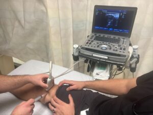
| Patient Preparation | – Positioning: Supine with knee extended (Image 1)
– Skin Preparation: Clean the knee with antiseptic solution (chlorhexidine or iodine) and drape the area. |
| Ultrasound Setup | – Equipment: Use a high-frequency linear probe (7.5-12 MHz) and portable ultrasound machine. A probe cover should be used.
– Probe Placement: Place the probe transversely over the knee, superior to the patella to visualize the suprapatellar pouch and effusion. Take note of the depth of the effusion as this will be needed prior to needle insertion (Image 1) |
| Needle Insertion | – Needle Selection: Typically a 20-25 gauge, 1.5-inch needle.
– Needle Placement: The needle should be inserted at a depth that corresponds to the depth of the effusion (Video 1, Image 1). For example, if the effusion is 2 cm deep, the needle should be inserted at a 2 cm depth, parallel to the joint line. This positioning will optimize needle visualization. Use real-time ultrasound to guide the needle into the suprapatellar space (Video 2). Avoid the quadriceps tendon and femoral condyles. |
| Injection of Medication | – Medications: Experts recommend injecting a mixture of a steroid, such as dexamethasone 4 mg, and an anesthetic, like bupivacaine 4-5 mL, into the knee effusion. This combination helps reduce inflammation and provides pain relief. Other common options include corticosteroids (e.g., triamcinolone), or local anesthetics (e.g., lidocaine, ropivacaine).
– Injection: Inject slowly after confirming the needle placement (Video 3). |
Video 1 Localization of suprapatellar effusion (Take note of the depth of the effusion, approximately 2 cm):
Video 2 Localization of needle entering the suprapatellar effusion:
Video 3 Injection of medications:
Post-Injection Assessment
After the injection, it is typical to observe for 15 minutes to assess for any immediate complications such as swelling, bleeding, or signs of infection.
Post-Procedure Care
After the injection, apply a sterile bandage or dressing to the site to minimize the risk of infection. Patients should be informed about potential side effects, including transient swelling, local infection, or in rare cases, knee joint infection (septic arthritis). Repeated corticosteroid and anesthetic injections may weaken joint structures over time, so it is important to limit their frequency—usually no more than an injection every 3-4 months—and monitor for long-term effects. While avascular necrosis is uncommon, repeated corticosteroid use has been associated with this condition in weight-bearing joints like the hip and knee. Regular follow-up is essential for long-term pain control and also to assess for any adverse effects, and patients should report any unusual pain or changes in mobility.
Conclusion
For chronic knee pain in the emergency department, ultrasound-guided suprapatellar knee injections provide an effective, minimally-invasive treatment option. By leveraging real-time imaging, emergency physicians can accurately deliver medications like corticosteroids or anesthetics to provide significant pain relief and improve function. Whether treating osteoarthritis, meniscal tears, or inflammatory arthritis, this technique is a valuable tool in managing chronic knee pain in the ED setting.
Key Points
- Ultrasound-guided suprapatellar knee injections are effective for chronic knee pain in the ED.
- Ultrasound improves accuracy, comfort, and outcomes compared to landmark-based injections.
- Indications include osteoarthritis, tendinopathy, inflammatory arthritis, trauma, post-surgical pain (with approval), and effusions.
- Contraindications include infection, coagulopathy, severe deformities, recent surgery, allergies, and certain medical conditions.
- Procedure: Supine position, antiseptic preparation, ultrasound guidance to insert a needle into the suprapatellar pouch and inject a steroid/anesthetic mix.
- Post-injection: Monitor for complications, educate on potential side effects, limit injection frequency, and ensure follow-up

Image 1: Knee position. Probe position.
References
- Saha P, Smith M, Hasan K. Accuracy of Intraarticular Injections: Blind vs. Image Guided Techniques-A Review of Literature. J Funct Morphol Kinesiol. 2023 Jun 29;8(3):93. doi: 10.3390/jfmk8030093. PMID: 37489306; PMCID: PMC10366715.
- Fang WH, Chen XT, Vangsness CT Jr. Ultrasound-Guided Knee Injections Are More Accurate Than Blind Injections: A Systematic Review of Randomized Controlled Trials. Arthrosc Sports Med Rehabil. 2021 Jun 26;3(4):e1177-e1187. doi: 10.1016/j.asmr.2021.01.028. PMID: 34430899; PMCID: PMC8365196.
- Steinbach LS, Stevens KJ. Imaging of cysts and bursae about the knee. Radiol Clin North Am. 2013 May;51(3):433-54. doi: 10.1016/j.rcl.2012.10.005. Epub 2013 Jan 12. PMID: 23622093.
- Wu T, Dong Y, Song Hx, Fu Y, Li JH. Ultrasound-guided versus landmark in knee arthrocentesis: A systematic review. Semin Arthritis Rheum. 2016 Apr;45(5):627-32. doi: 10.1016/j.semarthrit.2015.10.011. Epub 2015 Dec 17. PMID: 26791571.